Skip directly to site content Skip directly to page options Skip directly to A-Z link Skip directly to A-Z link Skip directly to A-Z link
Volume 21, Number 7—July 2015
Dispatch

Detection of Circovirus in Foxes with Meningoencephalitis, United Kingdom, 2009–2013

Steve Bexton, Lidewij C. Wiersma, Sarah Getu, Peter R. van Run, Georges M.G.M. Verjans, Debby Schipper, Claudia M.E. Schapendonk, Rogier Bodewes, Lucy Oldroyd, Bart L. Haagmans, Marion M.P. Koopmans, and Saskia L. SmitsComments to Author 
Author affiliations: RSPCA Norfolk Wildlife Hospital, East Winch, United Kingdom (S. Bexton); Erasmus Medical Center, Rotterdam, the Netherlands (L.C. Wiersma, S. Getu, P.R. van Run, G.M.G.M. Verjans, D. Schipper, C.M.E. Schapendonk, R. Bodewes, B.L. Haagmans, M.M.P. Koopmans, S.L. Smits); Abbey Veterinary Services, Newton Abbot, United Kingdom (L. Oldroyd); National Institute for Public Health and the Environment, Bilthoven, the Netherlands (M.M.P. Koopmans)

Main Article

Figure 1

Histopathologic features of brain tissue from foxes with possible virus-induced neurologic disease. A) Multifocal, randomly distributed areas of severe encephalitis and meningitis in the cerebrum (original magnification ×40). B) Detail of encephalitis in the cerebrum (original magnification ×200). Gray and, to a lesser extent, white matter of the cerebrum showed randomly dispersed areas of astrocytosis, gliosis, and infiltration with lymphocytes and plasma cells. Blood vessels in affected areas

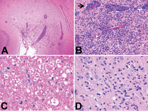
Figure 1. Histopathologic features of brain tissue from foxes with possible virus-induced neurologic disease. A) Multifocal, randomly distributed areas of severe encephalitis and meningitis in the cerebrum (original magnification ×40). B) Detail of encephalitis in the cerebrum (original magnification ×200). Gray and, to a lesser extent, white matter of the cerebrum showed randomly dispersed areas of astrocytosis, gliosis, and infiltration with lymphocytes and plasma cells. Blood vessels in affected areas show perivascular cuffing with distention of Virchow-Robin spaces with up to 10 layers of lymphocytes and plasma cells (arrow). C) Detail of white matter in the cerebellum (original magnification ×400). Axons in affected white matter showed degeneration, characterized by formation of spheroids, shrinkage, and fragmentation; axon sheaths containing microglia or macrophages; and presence of gitter cells in surrounding neuropil. Cerebellum was mildly affected, and meninges, especially of the cerebrum, were frequently distended with lymphocytes and plasma cells. D) Detail of gray matter of cerebrum (original magnification ×400). Individual neuronal cell bodies were frequently surrounded by up to 5 glial cells (i.e., sattelitosis) and showed margination of Nissl substance, hyperchromasia, degeneration, and necrosis. Tissue sections were subjected to conventional hematoxylin and eosin staining.

Main Article

Page created: June 15, 2015
Page updated: June 15, 2015
Page reviewed: June 15, 2015
The conclusions, findings, and opinions expressed by authors contributing to this journal do not necessarily reflect the official position of the U.S. Department of Health and Human Services, the Public Health Service, the Centers for Disease Control and Prevention, or the authors' affiliated institutions. Use of trade names is for identification only and does not imply endorsement by any of the groups named above.
file_external