Volume 22, Number 7—July 2016
Dispatch
Vesicular Disease in 9-Week-Old Pigs Experimentally Infected with Senecavirus A
Figure 1

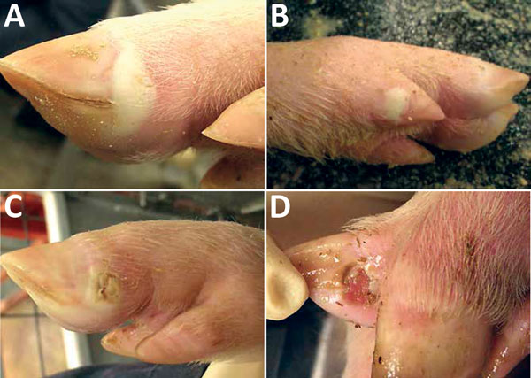
Figure 1. Vesicular lesions on feet of pigs experimentally infected with Senecavirus A. A) Blanched, intact, fluid-filled vesicle on lateral coronary band of toe. B) Intact vesicle on coronary band of medial dewclaw. C) Ruptured vesicle on coronary band of toe. D) Ruptured vesicle with ulceration and erosion in interdigital space.
1These authors contributed equally to this article.
Page created: June 14, 2016
Page updated: June 14, 2016
Page reviewed: June 14, 2016
The conclusions, findings, and opinions expressed by authors contributing to this journal do not necessarily reflect the official position of the U.S. Department of Health and Human Services, the Public Health Service, the Centers for Disease Control and Prevention, or the authors' affiliated institutions. Use of trade names is for identification only and does not imply endorsement by any of the groups named above.