Skip directly to site content Skip directly to page options Skip directly to A-Z link Skip directly to A-Z link Skip directly to A-Z link
Volume 22, Number 8—August 2016
Dispatch

Baylisascaris procyonis–Associated Meningoencephalitis in a Previously Healthy Adult, California, USA

Charles LangelierComments to Author , Michael J. Reid, Cathra Halabi, Natalie Witek, Alejandro LaRiviere, Maulik Shah, Michael R. Wilson, Peter Chin-Hong, Vanja Douglas, Kevin R. Kazacos, and Jennifer M. Babik
Author affiliations: University of California, San Francisco, California, USA (C. Langelier, M.J. Reid, C. Halabi, N. Witek, A. LaRiviere, M. Shah, M.R. Wilson, P. Chin-Hong, V. Douglas, J.M. Babik); Purdue University College of Veterinary Medicine, West Lafayette, Indiana, USA (K.R. Kazacos)

Main Article

Figure 2

Magnetic resonance imaging scans showing brain abnormalities in a previously healthy adult with Baylisascaris meningoencephalitis, California, USA. A–D) Postgadolinium contrast T1 images obtained 4 weeks after symptom onset. A–C) Axial images, moving inferiorly to superiorly, demonstrating nodular bilateral enhancement within the cerebellar hemispheres, thalami, and subcortical white matter. D) Sagittal image further demonstrates multifocal areas of enhancement in cerebral hemispheres. Additiona

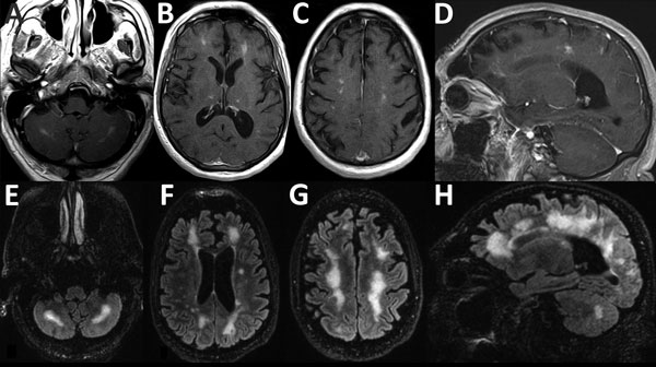
Figure 2. Magnetic resonance imaging scans showing brain abnormalities in a previously healthy adult with Baylisascaris meningoencephalitis, California, USA. A–D) Postgadolinium contrast T1 images obtained 4 weeks after symptom onset. A–C) Axial images, moving inferiorly to superiorly, demonstrating nodular bilateral enhancement within the cerebellar hemispheres, thalami, and subcortical white matter. D) Sagittal image further demonstrates multifocal areas of enhancement in cerebral hemispheres. Additional, mild T2 abnormalities (not shown) were present at the same time. E–H) T2/FLAIR (fluid attenuation inversion recovery) images obtained 6 weeks after symptom onset. E–G) Axial images, moving inferiorly to superiorly, demonstrating patchy and confluent hyperintense lesions throughout the supratentorial white matter and cerebellum. H) Sagittal image further demonstrates the high degree of white matter abnormality, which was not present on the earlier imaging. Postcontrast enhancement on T1 imaging (not shown) had nearly resolved at this time.

Main Article

Page created: September 12, 2016
Page updated: September 12, 2016
Page reviewed: September 12, 2016
The conclusions, findings, and opinions expressed by authors contributing to this journal do not necessarily reflect the official position of the U.S. Department of Health and Human Services, the Public Health Service, the Centers for Disease Control and Prevention, or the authors' affiliated institutions. Use of trade names is for identification only and does not imply endorsement by any of the groups named above.
file_external