Volume 6, Number 4—August 2000
Dispatch
Subacute Sclerosing Panencephalitis, a Measles Complication, in an Internationally Adopted Child
Figure 2
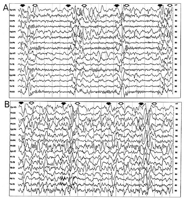
Figure 2. Electroencephalogram (EEG) at the time of presentation in the neurology clinic (A) and 3 months later (B). The initial EEG (A) reveals periodic bursts of high-amplitude, slow-wave complexes. (Onset of the complexes is indicated by solid arrows; offset, by open arrows.) The background rhythm is normal, except for bifrontal slowing. This "burst-suppression" pattern is highly characteristic of subacute sclerosing panencephalitis (4). EEG 3 months later, when the patient's clinical status has worsened (B), again shows periodic high-amplitude slow waves (again, between the solid and open arrows), but they now arise from a diffusely slowed background rhythm, which nearly obscures the periodic slow waves. In both A and B, the interval between each vertical dotted line is one second.
References
- Hostetter MK. Infectious diseases in internationally adopted children: the past five years. Pediatr Infect Dis. 1998;17:517–8. DOIGoogle Scholar
- Hostetter MK. Infectious diseases in internationally adopted children: Findings in children from China, Russia, and Eastern Europe. Adv Pediatr Infect Dis. 1999;14:147–61.PubMedGoogle Scholar
- Gascon GG. Subacute sclerosing panencephalitis. Semin Pediatr Neurol. 1996;3:260–9. DOIPubMedGoogle Scholar
- Britt WJ. Slow viruses. In: Feigin R, Cherry J, editors. Textbook of pediatric infectious diseases. 4th ed. Philadelphia: W.B. Saunders Company, 1998; p. 646-65.
- Dyken PR. Subacute sclerosing panencephalitis: current status. Neurol Clin. 1985;3:179–96.PubMedGoogle Scholar
- Norrby E, Kristensson K. Measles virus in the brain. Brain Res Bull. 1997;44:213–20. DOIPubMedGoogle Scholar
- PeBenito R, Naqvi SH, Arca MM, Schubert R. Fulminating subacute sclerosing panencephalitis: Case report and literature review. Clin Pediatr. 1997;36:149–54. DOIGoogle Scholar
- Crowell J. Clinical neurophysiology: Video EEG studies. Symposium to ICNA/CNS Subacute Sclerosing Panencephalitis meeting: Update. San Francisco, CA, Oct 1, 1994.
- Winer JB, Pires M, Kernode A, Ginsberg L, Rosser M. Resolving MRI abnormalities with progression of SSPE. Neuroradiology. 1991;33:178–80. DOIPubMedGoogle Scholar
- Anlar B, Saatci I, Kose G, Yalaz K. MRI findings in subacute sclerosing panencephalitis. Neurology. 1996;47:1278–83.PubMedGoogle Scholar
- Geller TJ, Vern BA, Sarwar M. Focal MRI findings in early SSPE. Pediatr Neurol. 1987;3:310–2. DOIPubMedGoogle Scholar
- Park SY. Subacute sclerosing panencephalitis in an identical twin. Pediatrics. 1999;104:1390–4. DOIPubMedGoogle Scholar
- Notifiable diseases/deaths in selected cities. MMWR Morb Mortal Wkly Rep. 2000;48:1183–90.
- Moodley M. Subacute sclerosing panencephalitis in the developing world. S Afr Med J. 1992;82:72–3.PubMedGoogle Scholar