Volume 9, Number 12—December 2003
Research
Mycobacterium abscessus and Children with Cystic Fibrosis
Cite This Article
Citation for Media
Abstract
We prospectively studied 298 patients with cystic fibrosis (mean age 11.3 years; range 2 months to 32 years; sex ratio, 0.47) for nontuberculous mycobacteria in respiratory samples from January 1, 1996, to December 31, 1999. Mycobacterium abscessus was by far the most prevalent nontuberculous mycobacterium: 15 patients (6 male, 9 female; mean age 11.9 years; range 2.5–22 years) had at least one positive sample for this microorganism (versus 6 patients positive for M. avium complex), including 10 with >3 positive samples (versus 3 patients for M. avium complex). The M. abscessus isolates from 14 patients were typed by pulsed-field gel electrophoresis: each of the 14 patients harbored a unique strain, ruling out a common environmental reservoir or person-to-person transmission. Water samples collected in the cystic fibrosis center were negative for M. abscessus. This major mycobacterial pathogen in children and teenagers with cystic fibrosis does not appear to be acquired nosocomially.
Since 1990, an increasing number of studies have reported the recovery of nontuberculous mycobacteria from the respiratory tract of patients with cystic fibrosis (1–4). Mycobacterium abscessus (formerly M. chelonae subsp. abscessus), a rapidly growing mycobacterium of the M. fortuitum complex, is of particular concern. It can cause severe lung disease, which spontaneously advances until it becomes debilitating or fatal (5,6). It may be responsible for disseminated infections in patients undergoing lung transplantation (7). This organism is usually also susceptible to only a few drugs (8), and some strains may exhibit multidrug resistance (7).
The frequency of isolation of M. abscessus in cystic fibrosis patients is unclear. Many studies on nontuberculous mycobacteria in such patients did not distinguish M. abscessus and M. chelonae, formerly two subspecies of M. chelonae, and used the designation M. chelonae–M. abscessus, M. chelonae group, or even M. fortuitum complex. Moreover, most studies were conducted with adults (1,2,4). How cystic fibrosis patients become contaminated is also poorly understood. M. abscessus has been reported to be acquired iatrogenically in non–cystic fibrosis patients (9). The members of the M. fortuitum complex are saprophytic organisms living in soil and water that are ubiquitous in hospital environments and survive well in adverse conditions (10–13). Aerosols, pulmonary function equipment, and bronchoscopes are thus potential sources of contamination for patients with cystic fibrosis. Alternatively, transmission from patients to patients attending the same department-care facilities might occur, although this finding has been recently challenged (14).
We encountered one case of M. abscessus infection in a patient with cystic fibrosis in 1995. The recovery of this unusual pathogen prompted us to prospectively evaluate the rate of isolation of M. abscessus in the cystic fibrosis patients attending our center, the degree of transmissibility of this organism, and its clonality, by using DNA-based identification and typing systems.
Description of Study
All patients with cystic fibrosis who attended the pediatric department of Necker Hospital for Sick Children from January 1, 1996, to December 31, 1999, provided at least one sputum sample per year, which was processed for the culture of acid-fast bacilli (AFB). Patients who provided a positive sample then submitted >3 sputum samples for AFB smear and culture over the next 3 months. AFB smears and cultures were checked quarterly thereafter.
Cultures of Respiratory Specimens
Specimens were decontaminated with NALC-NaOH-oxalic acid (0.25% N-acetyl-L-cysteine–1% sodium hydroxide–5% oxalic acid) (15). AFB smears were stained with auramine-rhodamine and scored as previously described (16). Two Löwenstein-Jensen slants were inoculated for each specimen, one of which was incubated at 37°C and the other at 30°C. The slants were examined twice weekly for 2 weeks and then weekly for a further 10 weeks.
Environmental Samples
Water samples taken from the hospital’s hot and cold water supply systems were collected in sterile plastic bottles. Samples (100 mL) were decontaminated with 1% NaOH without prior concentration by filtration (17). The inner surfaces of respiratory devices (e.g., nebulizers, bronchoscopes) were rinsed with 1 to 10 mL of sterile distilled water; the water used for rinsing was processed for the culture of AFB without prior decontamination with 1% NaOH.
Species Identification
Rapidly growing mycobacteria recovered from clinical and environmental samples were identified by standard techniques (17) and hsp65 sequencing (18). The M. avium complex was identified by the AccuProbe technique (Gen-Probe Inc., San Diego, CA). The hsp65 genomovars of M. abscessus were referred to as T (identical to the type-strain M. abscessus ATCC 19977T), -5a (differing from ATCC 19977T by 5 nt, and identical to the reference strain M. abscessus IP970272), -5b (differing from ATCC 19977T by 5 nt, and identical to the reference strain M. abscessus IP970453), and –6 (differing from ATCC 19977T by 6 nt, and identical to the reference strain M. abscessus IP140420009), as previously described (18).
PFGE Analysis
M. abscessus isolates were analyzed by PFGE as described by Wallace et al., with minor modifications (9). Restriction fragments obtained after digestion with DraI and XbaI were separated in 0.5 x TBE buffer (0.025 M Tris, 0.5 mM EDTA, and 0.025 M boric acid) supplemented with 50 μM thiourea (19), using a CHEF-DR III system (Bio-Rad, Richmond, CA) at 14°C and 6 V/cm2. Pulse times were ramped linearly from 1.5 to 21.5 s for 23 h. A size standard (bacteriophage λ concatemers) was run in parallel in each experiment. Restriction patterns were analyzed with the Taxotron package (Taxolab Software, Institut Pasteur, Paris, France) comprising the RestrictoScan, RestrictoTyper, Adanson, and Dendrograph programs.
Screening the Study Population for Nontuberculous Mycobacteria
A total of 298 patients with cystic fibrosis (1,525 sputum samples; mean of 5.0 samples per patient) followed up at our institution were screened for M. abscessus from January l, 1996, to December 31, 1999. The age of the patients ranged from 2 months to 32 years (mean 11.3 years). The sex ratio was 0.47 (140 male/158 female patients). Samples from two patients could not be analyzed because the samples were repeatedly contaminated (<1 interpretable culture per year during the study period). Of the 296 patients with interpretable cultures, 29 (9.80%) provided at least one sample positive for nontuberculous mycobacteria. Twelve of the 296 patients had M. abscessus alone, 3 had M. abscessus and M. gordonae, 6 had M. avium complex, 4 had M. gordonae, 1 had M. fortuitum, 1 had M. kansasii, and 2 had organisms not related to any known species. Thirteen patients provided at least three positive samples, 10 involving M. abscessus and 3 M. avium complex. Two of these patients were twin sisters, one colonized with M. abscessus (patient no. 5) and the other with M. avium complex.
Description of Cases with M. abscessus Isolation
Fifteen (5%) of the 296 patients with interpretable cultures provided at least one sample positive for M. abscessus. Ten of these patients had >3 positive samples, including six with positive AFB smears (Table). Mycobacterial disease was documented in four patients: a 16-year-old boy (patient no. 4) with parenchymal condensation of the left lower lobe on chest x-ray and computed tomographic (CT) scan, which disappeared only under anti–M. abscessus treatment; a 10-year-old boy (patient no. 6), whose rapidly deteriorating and ultimately fatal condiction was associated with diffuse bronchiectasis on CT scan; a 9-year-old girl (patient no. 7), who had a massive, granulomatous pneumonia of the right lung that led to pneumonectomy, and who died after 15 months of bacteriologically ineffective anti–M. abscessus treatment; and a 2-1/2-year-old girl (patient no. 8) with segmental condensation of the right mid-lobe on chest x-ray and CT scan, which disappeared only under anti–M. abscessus treatment.
All but one (patient no. 1) of the 15 patients were recognized during the study period. Some of the patients who were identified in the first year may have previously gone undetected, as nontuberculous mycobacteria had not been sought before (patients nos. 2, 3, 4, 5, and 7). The M. abscessus isolates belonged to genomovars T, -5a, and -6, with a slightly higher prevalence of genomovar T (Table). Genomovar T was involved in the two fatal cases recorded during the study period (patients nos. 6 and 7).
Characteristics of Patients Positive for M. abscessus
The 15 patients positive for M. abscessus were predominantly females (sex ratio, 0.40). Their mean age at the time of the first culture positive for M. abscessus (11.9 years, range 2.5–22 years) was very similar to the mean age of the entire study population. However, the mean age was lower than that for patients positive for M. avium complex (17.5 years; range 13–25 years). Of the 14 patients who underwent genotype analysis, 8 were homozygous for deletion of the phenylalanine in position 508, and 4 were heterozygotous for this deletion plus another mutation. Pulmonary function at the time of the first isolation of M. abscessus was highly variable, with forced expiratory volume in 1 second and forced vital capacity values ranging from 14% to 99%, and 31% to 104% of predicted values, respectively. Schwachman score (20) also greatly varied among patients (range 40–85). The most prevalent associated disorders included bronchiectasis (13 cases), gastroesophageal reflux (3 cases), and allergic bronchopulmonary aspergillosis (3 cases). All patients had pancreatic insufficiency. Nine of the 15 patients were colonized (at least three positive sputum samples within the previous 12 months) with Pseudomonas aeruginosa. None was colonized with Burkholderia cepacia.
We analyzed records of all treatments received by the patients within the 12 months preceding the first isolation of M. abscessus, including therapeutic aerosols. All of the patients had received IV antibiotics (1–5 two-week IV courses; median 3 courses), combined with aerosol antibiotics at home in 11 patients (tobramycin, 3 patients; colistin, 8 patients). Six patients had received aerosolized deoxyribonuclease. Two patients had received oral corticosteroids, and four had received inhaled corticosteroids.
Environmental Study
A total of 93 water samples collected from 40 water supply points in the cystic fibrosis center were studied. Three samples (3.2%) from two water supply points tested positive for rapidly growing mycobacteriua (M. mucogenicum, two samples; M. peregrinum: one sample). None of the samples tested positive for M. abscessus. None of the 12 respiratory devices (3 bronchoscopes, 9 nebulizers) studied in October 1997 tested positive for any nontuberculous mycobacteria.
PFGE Analysis of M. abscessus Isolates
Figure
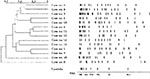
Figure. Pulsed-field gel electrophoresis analysis of DraI-digested DNA from Mycobacterium abscessus isolates. Restriction patterns of isolates from 14 patients are shown, with a dendrogram of similarity; λ concatemers were used as size standards.
PFGE was used to compare the isolates from 14 patients positive for M. abscessus (the isolates from patient no. 7 could not be subcultured for testing because of inadequate storage). We studied all isolates from each patient who provided <3 positive cultures and a maximum of five isolates from each patient with >3 isolates. The isolates from three patients (patients nos. 6, 11, and 12) gave unreadable restriction patterns with classical protocols, despite multiple attempts. This phenomenon is common with mycobacteria, particularly M. abscessus (9) and is probably related to Tris-dependent site-specific cleavage of the DNA (19). Nondegradative PFGE was only achieved by running gels in the presence of thiourea, which has been shown to protect the DNA from strand cleavage (21). We were therefore able to type all isolates from the 14 patients by PFGE. Each of these 14 patients had isolates of a unique genotype that was unrelated to the genotype of any other patient (Figure). No differences were detected between isolates from the same patient (data not shown), even if the interval between the first and last isolation was as long as 4 years (patient no. 1).
M. abscessus was the predominant nontuberculous mycobacterium recovered from the patients attending our center. Approximately 5% of the patientswe screened provided at least one sputum sample positive for this organism, and >65% of these patients had >3 positive samples. Other rapidly growing mycobacteria were far behind (M. fortuitum, one patient). This finding confirms that M. abscessus differs from M. chelonae and from other members of the M. fortuitum complex by its particular propensity to cause lung disease in a variety of clinical settings. In a series of 154 cases of lung infection caused by rapidly growing mycobacterium in patients with and without cystic fibrosis, >80% of isolates were M. abscessus; M. fortuitum was isolated in <15% and M. chelonae in <1% of patients (22).
In contrast with other studies on cystic fibrosis populations composed of teenagers and adults (2,4), we found that the M. avium complex was isolated less frequently, with an overall prevalence (percentage of patients with at least one positive nontuberculous mycobacterial culture) of approximately 2%. Other pediatric cystic fibrosis centers have reported similar findings (23,24). This finding suggests that M. abscessus is the most prevalent mycobacterial pathogen in children and teenagers with cystic fibrosis. This finding is further supported by the lack of cases involving M. avium complex in patients <13 years of age. However, this finding does not preclude epidemiologic variations between countries or institutions.
Nosocomial acquisition of M. abscessus has been well documented in patients without cystic fibrosis. Several nosocomial outbreaks of infection or pseudoinfection have been attributed to this organism after cardiac surgery, bacteremia associated with hemodialysis, and pseudoinfections due to contaminated bronchoscopes (9). Epidemiologic investigations showed that these outbreaks resulted from the use of contaminated water. Disinfectants may be ineffective against M. abscessus in real conditions of use (12). PFGE has been used to retrospectively analyze the clinical and environmental isolates recovered during M. abscessus outbreaks (9). Each of the outbreaks with typeable isolates clearly involved a single strain, which was usually recovered from the water supply system.
Our results do not support nosocomial acquisition of M. abscessus. First, the recovery rate of this organism remained constantly low throughout the study. Previous nosocomial outbreaks involving patients without cystic fibrosis were characterized by much higher attack rates. Second, we did not find any link between the use of respiratory devices and the acquisition of M. abscessus. Patients positive for M. abscessus did not receive more aerosol treatments than did patients with similar clinical status (not shown). The patients used their personal nebulizer at the center and received aerosols in their own rooms. Sterile saline was used when the aerosol was mixed. Reusable respiratory devices were disinfected according to validated protocols and were washed exclusively with sterile water. During the study period, no patients without cystic fibrosis, even severely immunocompromised ones, were infected with M. abscessus as a result of a contaminated bronchoscope in our pediatric department. Third, although various rapidly growing mycobacteria were recovered from several water supply points in our center, M. abscessus was not isolated. Finally, PFGE analysis demonstrated that the cases involved unrelated strains, which argues against a common source of contamination or patient-to-patient transmission. Similar results have been recently reported with fewer patients (14). The low transmissibility, if any, of M. abscessus from person to person is further supported by the observation of twin sisters in our series, only one of whom was colonized with M. abscessus.
Whether specific measures are necessary to prevent M. abscessus infection in patients with cystic fibrosis is questionable (14). Our epidemiologic results indicate few potential control approaches exist. A strict segregation policy seems unnecessary because apparently no risk of person-to-person transmission of M. abscessus exists (14, this study). Further epidemiologic studies are required before recommendations for infection-control precautions can be formulated.
Dr. Sermet-Gaudelus is a physician in the Pediatric Department of the Necker Hospital for Sick Children, where she is studying a cohort of 350 cystic fibrosis patients. She is especially interested in emerging bacterial and fungal agents in such patients. She is now finishing a doctoral thesis on the electrophysiology of the nasal mucosa in cystic fibrosis patients.
Acknowledgments
We thank Gilles Quesne, Maria-Cristina Guttierez-Perez, and Martin Rottman for their contribution to this work.
This work received financial support from the Association “Vaincre la Mucoviscidose.”
References
- Kilby JM, Gilligan PH, Yankaskas JR, Highsmith WE Jr, Edwards LJ, Knowles MR. Nontuberculous mycobacteria in adult patients with cystic fibrosis. Chest. 1992;102:70–5. DOIPubMedGoogle Scholar
- Aitken ML, Burke W, McDonald G, Wallis C, Ramsey B, Nolan C. Nontuberculous mycobacterial disease in adult cystic fibrosis patients. Chest. 1993;103:1096–9. DOIPubMedGoogle Scholar
- Hjelt K, Hojlyng N, Howitz P, Illum N, Munk E, Valerius NH, The role of mycobacteria other than tuberculosis (MOTT) in patients with cystic fibrosis. Scand J Infect Dis. 1994;26:569–76. DOIPubMedGoogle Scholar
- Olivier KN, Yankaskas JR, Knowles MR. Nontuberculous mycobacterial pulmonary disease in cystic fibrosis. Semin Respir Infect. 1996;11:272–84.PubMedGoogle Scholar
- Tomashefski JF Jr, Stern RC, Demko CA, Doershuk CF. Nontuberculous mycobacteria in cystic fibrosis. An autopsy study. Am J Respir Crit Care Med. 1996;154:523–8.PubMedGoogle Scholar
- Cullen N, Cannon CL, Mark EJ, Colin AA. Mycobacterium abscessus infection in cystic fibrosis: colonization or infection. Am J Respir Crit Care Med. 2000;161:641–5.PubMedGoogle Scholar
- Sanguinetti M, Ardito F, Fiscarelli E, La Sorda M, D’Argenio P, Ricciotti G, Fatal pulmonary infection due to multidrug-resistant Mycobacterium abscessus in a patient with cystic fibrosis. J Clin Microbiol. 2001;39:816–9. DOIPubMedGoogle Scholar
- American Thoracic Society. Diagnosis and treatment of disease caused by nontuberculous mycobacteria. Medical Section of the American Lung Association. Am J Respir Crit Care Med. 1997;156:S1–25.PubMedGoogle Scholar
- Wallace RJ Jr, Zhang Y, Brown BA, Fraser V, Mazurek GH, Maloney S. DNA large restriction fragment patterns of sporadic and epidemic nosocomial strains of Mycobacterium chelonae and Mycobacterium abscessus. J Clin Microbiol. 1993;31:2697–701.PubMedGoogle Scholar
- Carson LA, Petersen NJ, Favero MS, Aguero SM. Growth characteristics of atypical mycobacteria in water and their comparative resistance to disinfectants. Appl Environ Microbiol. 1978;36:839–46.PubMedGoogle Scholar
- Carson LA, Bland LA, Cusick LB, Favero MS, Bolan GA, Reingold AL, Prevalence of nontuberculous mycobacteria in water supplies of hemodialysis centers. Appl Environ Microbiol. 1988;54:3122–5.PubMedGoogle Scholar
- Lowry PW, Beck-Sague CM, Bland LA, Aguero SM, Arduino MJ, Minuth AN, Mycobacterium chelonae infection among patients receiving high-flux dialysis in a hemodialysis clinic in California. J Infect Dis. 1990;161:85–90.PubMedGoogle Scholar
- Falkinham JO III. Epidemiology of infection by nontuberculous mycobacteria. Clin Microbiol Rev. 1996;9:177–215.PubMedGoogle Scholar
- Bange FC, Brown BA, Smaczny C, Wallace RJ Jr, Bottger EC. Lack of transmission of Mycobacterium abscessus among patients with cystic fibrosis attending a single clinic. Clin Infect Dis. 2001;32:1648–50. DOIPubMedGoogle Scholar
- Whittier S, Hopfer RL, Knowles MR, Gilligan PH. Improved recovery of mycobacteria from respiratory secretions of patients with cystic fibrosis. J Clin Microbiol. 1993;31:861–4.PubMedGoogle Scholar
- Whittier S, Olivier K, Gilligan P, Knowles M, Della-Latta P. The nontuberculous mycobacteria in cystic fibrosis study group. Proficiency testing of clinical microbiology laboratories using modified decontamination procedures for detection of nontuberculous mycobacteria in sputum samples from cystic fibrosis. J Clin Microbiol. 1997;35:2706–8.PubMedGoogle Scholar
- Nolte FS, Metchock B. Mycobacterium. In: Baron EJ, Murray PR, Pflaller MA, Tenover FC, Yolken RH, editors. Manual of clinical microbiology. 6th ed. Washington: American Society for Microbiology; 1995. p. 400–37.
- Ringuet H, Akoua-Koffi C, Honore S, Varnerot A, Vincent V, Berche P, hsp65 sequencing for identification of rapidly growing mycobacteria. J Clin Microbiol. 1999;37:852–7.PubMedGoogle Scholar
- Galamba A, Soetaert K, Wang XM, De Bruyn J, Jacobs P, Content J. Disruption of adhC reveals a large duplication in the Mycobacterium smegmatis mc(2)155 genome. Microbiology. 2001;147:3281–94.PubMedGoogle Scholar
- Schwachman H, Kulczycki L. Long term study of 105 patients with cystic fibrosis. Am J Dis Child. 1958;96:6–15.
- Evans M, Dyson P. Pulsed-field gel electrophoresis of Streptomyces lividans DNA. Trends Genet. 1993;9:72. DOIPubMedGoogle Scholar
- Griffith DE, Girard WM, Wallace RJ Jr. Clinical features of pulmonary disease caused by rapidly growing mycobacteria. An analysis of 154 patients. Am Rev Respir Dis. 1993;147:1271–8.PubMedGoogle Scholar
- Boxerbaum B. Isolation of rapidly growing mycobacteria in patients with cystic fibrosis. J Pediatr. 1980;96:689–91. DOIPubMedGoogle Scholar
- Fauroux B, Delaisi B, Clement A, Saizou C, Moissenet D, Truffot-Pernot C, Mycobacterial lung disease in cystic fibrosis: a prospective study. Pediatr Infect Dis J. 1997;16:354–8. DOIPubMedGoogle Scholar
Figure
Table
Cite This ArticleTable of Contents – Volume 9, Number 12—December 2003
| EID Search Options |
|---|
|
|
|
|
|
|
Please use the form below to submit correspondence to the authors or contact them at the following address:
Jean-Louis Gaillard, Laboratoire de Microbiologie, Hôpital Raymond Poincaré, 104 Boulevard Raymond Poincaré, 92380, Garches, France; fax: +33 147 10 79 49
Top