Volume 9, Number 8—August 2003
Research
Community-Acquired Methicillin-Resistant Staphylococcus aureus Carrying Panton-Valentine Leukocidin Genes: Worldwide Emergence
Cite This Article
Citation for Media
Abstract
Infections caused by community-acquired (CA)-methicillin resistant Staphylococcus aureus (MRSA) have been reported worldwide. We assessed whether any common genetic markers existed among 117 CA-MRSA isolates from the United States, France, Switzerland, Australia, New Zealand, and Western Samoa by performing polymerase chain reaction for 24 virulence factors and the methicillin-resistance determinant. The genetic background of the strain was analyzed by pulsed-field gel electrophoresis (PFGE) and multi-locus sequence typing (MLST). The CA-MRSA strains shared a type IV SCCmec cassette and the Panton-Valentine leukocidin locus, whereas the distribution of the other toxin genes was quite specific to the strains from each continent. PFGE and MLST analysis indicated distinct genetic backgrounds associated with each geographic origin, although predominantly restricted to the agr3 background. Within each continent, the genetic background of CA-MRSA strains did not correspond to that of the hospital-acquired MRSA.
Methicillin-resistant Staphylococcus aureus (MRSA) are identified as nosocomial pathogens throughout the world (1). Established risk factors for MRSA infection include recent hospitalization or surgery, residence in a long-term–care facility, dialysis, and indwelling percutaneous medical devices and catheters. Recently, however, cases of MRSA have been documented in healthy community-dwelling persons without established risk factors for MRSA acquisition. Because they are apparently acquired in the community, these infections are referred to as community-acquired (CA)-MRSA (2). CA-MRSA infections have been reported in North America, Europe, Australia, and New Zealand (3–5). The recent genomic sequence of a CA-MRSA isolate (6) indicated the presence not only of a novel smaller variant of the methicillin-resistance locus (SCCmec IVa, according to Baba et al. designation [6]), but also that of the locus for the Panton-Valentine leukocidin (PVL). The PVL locus is carried on a bacteriophage and is present in only a small percentage of S. aureus isolates from France, where this locus is associated with skin infections, and occasionally, severe necrotizing pneumonia (7,8).
In a recent study, we found that CA-MRSA infections in France are caused by a single clone producing the PVL (3). Analysis of a set of CA-MRSA strains from the United States and Australia confirmed the presence of SCCmec IVa in most of them, and genetic comparison of the CA-MRSA by multi-locus sequence typing (MLST) indicated that they belonged to five clonal complexes, two of which predominated (4). This finding suggested that CA-MRSA have arisen from diverse genetic backgrounds rather than the worldwide spread of a single clone (4).
The aim of this study was to determine whether the PVL gene represents a stable marker of the CA-MRSA strains worldwide and whether any other common genetic traits such as toxin gene and the accessory gene regulator (agr) profiles can be identified.
Bacterial Strains
A total of 117 different isolates of CA-MRSA were examined. Community acquisition was defined as growth of the isolates within 48 hours after hospital admission in patients who had no risk factors for nosocomial acquisition, including no hospitalizations or nursing home residence in the year before admission. Thirty-three isolates were from the United States. The U.S. isolates all belonged to the previously identified major CA-MRSA clonal group and included strain MW2 (2). They originated from 12 different facilities in Minnesota; two isolates were from North Dakota. Sixty-seven isolates originated from Europe (61 isolates from France and 6 from Switzerland). The French CA-MRSA isolates originated from 10 different hospitals located throughout France, and the Swiss isolates were from the Geneva area. Seventeen isolates were from Oceania and included two different clones: 1) 13 isolates corresponded to the Western Samoan phage pattern strains, also designated the Southwest Pacific clone (9) and were isolated from Australia (8 isolates), New Zealand (4 isolates), and Western Samoa (1 isolate) (5); 2) 4 other isolates from Australia belonged to a recently described clone designated as the Queensland clone (10). The Southwest Pacific clone has two distinct phage-typing patterns known as Western Samoan Phage Pattern (WSPP) 1 and 2 (5). The Queensland clone, which cannot be phage typed, was first detected in the southeast Queensland city of Ipswich in 2000 (10). Most isolates were from primary skin and soft tissue infections, with some cases of bacteriemia and at least five cases of necrotizing pneumonia (2,3,9,11,12).
A set of representative hospital-acquired (HA)MRSA isolates was included in the study: 24 from France and 33 from the United States. French HA-MRSA isolates corresponded to those of the major clones isolated throughout France as described by Lelievre et al. (13). HA-MRSA isolates were recovered from the same 12 different facilities that CA-MRSA. HA-MRSA from the United States originated from the same geographic area as the CA-MRSA.
Antimicrobial Susceptibility Testing
The MICs of benzyl-penicillin, oxacillin, gentamicin, tobramycin, kanamycin, chloramphenicol, tetracycline, minocycline, erythromycin, lincomycin, pristinamycin, fusidic acid, rifampicin, ofloxacin, co-trimoxazole, linezolid, mupirocin, vancomycin, and teicoplanin were determined for selected isolates (46 European, 22 U.S., and 13 Oceanian isolates) by using the standardized agar dilution technique as recommended by the French Society for Microbiology (14).
Detection of Accessory Genes by PCR
Using polymerase chain reaction (PCR), we determined the presence of accessory gene regulator (agr) allele group (1–4), SCCmec element (I-IV, according to the designation of Oliveira [15]), and 22 specific staphylococcal virulence genes (including 16 super-antigenic toxins, 3 hemolysins, and 3 leukocidins), as described previously (16).
Amplification of gyrA was used as a quality control of each DNA extract and the absence of PCR inhibitors. S. aureus strains Fri 913 (sea, see, sec, tst, lukE lukD, sek, sel, sep, and hlg), Fri 1151m (sed, sej, lukE lukD, hlgv, and hlb), ATCC 14458 (CCM5757) (seb, lukE lukD, sek, and hlgv), NCTC 7428 (sec, tst, lukM, seg, sei, sem, sen, seo, lukE lukD, hlgv, and hlb), A92 0211 (seg, sei, sem, sen, seo, eta, etb, lukE lukD, and hlgv), RN6390 (lukE lukD, hlgv, hlb, and agr1), RN6607 (sed, seg, sei, sem, sen, seo, lukE lukD, hlgv, and agr2), RN8465 (seg, sei, sem, sen, seo, tst, hlg, and agr3), RN4850 (seg, sei, sem, sen, seo, eta, etb, lukE lukD, hlgv, and agr4), RN 6911(lukE lukD, hlgv, hlb, agr null), E-1 (seg, sei, sem, sen, seo, lukE lukD, eta, hlgv, edinB and C), ATCC 49775 (seg, sei, sem, sen, seo, lukS lukF, and hlg), and ATCC 51811 (FRI 569) (seh, lukE lukD, hlb, and hlgv) were used as positive controls for PCR (17,18). S. aureus COL (SCCmec I), PER34 (SCCmec IA), BK2464 (SCCmec II), ANS46 (SCCmec III), HU25 (SCCmec IIIA), and HDE288 (SCCmec IV) were used as controls for characterization of the mec element according to Oliveira and de Lencastre (15).
The overall genetic background of the isolates was evaluated: 1) by digesting whole cell DNA with SmaI macrorestriction enzyme and determining the fragment-size patterns obtained on pulsed-field gel electrophoresis (PFGE) using a contour-clamped homogeneous electric field system on a CHEF DR-II apparatus (Bio-Rad Laboratories, Marnes-la-Coquette, France) as previously described (19). Resolved macrorestriction patterns were compared as recommended by Tenover et al. (20). Isolates differing by up to three fragments were considered as subtypes of a given clonal type. MLST was performed as described by Enright et al. (21). Briefly, seven housekeeping genes were used in the scheme; for each isolate, the alleles at each of the seven loci defined the allelic profile, which corresponded to a sequence type (ST). ST designations were those assigned by the MLST database (available from: URL: http://www.mlst.net).
Distribution of Accessory Genes from MRSA in Three Continents
Overall, we detected 12 different virulence genes or gene clusters among the 117 isolates (Table 1). Two gene loci were common to CA-MRSA isolates from all locations. Methicillin resistance was conferred in all 117 CA-MRSA isolates by the truncated SCCmec type IV element, and all the isolates contained the PVL locus. In addition, 112 isolates harbored the related lukE-lukD genes of another leukocidin frequently recovered from patients with all types of staphylococcal infections (3). Most (113 [97%] of 117) isolates were of agr type 3.
The distribution of other genes varied by the continent of origin. The European and Southwest Pacific isolates had consistent, relatively simple, patterns of virulence-associated genes. In addition to the SCCmec type IV element, PVL genes, and lukE-lukD, all the European isolates were positive for hlg-v (the γ-hemolysin variant gene) and the Southwest Pacific isolates were positive for hlg (the γ-hemolysin gene) and egc (the enterotoxin gene cluster coding for the enterotoxins seg, sei, sem, sen, and seo) (Table 1). The Queensland isolates had not been tested for the toxin genes, except the PVL locus. In contrast, considerable variability existed among the 33 U.S. isolates. As for the European isolates, the U.S. isolates showed both the presence of hlg-v and the absence of hlg. However, seven other toxins (the enterotoxins sea–sed, sej, she, and sek), which were absent in non–U.S. isolates, were variously found in up to 29 (3% to 87%) of these 33 isolates (Table 1).
Analysis of Genetic Background of CA-MRSA by PFGE
Figure
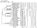
Figure. Pulsed-field gel electrophoresis (PFGE) pattern and phylogenetic tree of 117 community-acquired (CA)-methicillin resistant Staphylococcus aureus isolates from three continents. SmaI macrorestriction patterns were digitized and analyzed by using Taxotron software (Institut...
The 117 CA-MRSA isolates clustered into six PFGE clonal types (A to F, Figure). All 67 European isolates grouped into seven related PFGE patterns (subtypes A1-7) distinct from the other isolates. Most of the U.S. isolates belonged to a closely related group of five patterns (subtypes B1-5), although two well-differentiated outliers existed, one (D1) containing three isolates and the other (F1) containing only one isolate, both of which had an agr type 1 genotype. The 13 isolates obtained from Australia, New Zealand, and Western Samoa and belonging to the Southwest Pacific clone showed three closely related PFGE patterns (subtypes C1-3) with no geographic association. The four isolates from Australia belonging to the Queensland clone had the same E1 pattern. The phylogenetic tree shown on the left side of the Figure confirms the diversity in PFGE patterns between the CA-MRSA from different continents.
CA-MRSA Antibiotic Susceptibility Profiles
MICs were determined on a selected number of isolates (81) that corresponded to the different PFGE patterns. Overall, CA-MRSA isolates from all locations were susceptible to numerous antimicrobial drugs, including tobramycin, gentamicin, lincomycin, pristinamycin, minocycline, chloramphenicol, ofloxacin, vancomycin, teicoplanin, fosfomycin, rifampin, co-trimoxazole, and linezolid (Table 2). For the purposes of assessing differences between locations, isolates from the United States and Oceania were combined since their susceptibility profiles were similar (Table 2). Minor heterogeneity in erythromycin and mupirocin susceptibility was noted among European isolates. In contrast, U.S. and Oceanian isolates were uniformly susceptible to these antibiotics (Table 2). The main difference between the two groups was their susceptibility to kanamycin, tetracycline, and fusidic acid, with the European group of isolates being more resistant than the U.S. and Oceanian group (Table 2).
Comparison of CA-MRSA with HA-MRSA
Using PFGE to compare CA-MRSA isolates with representative HA-MRSA isolates from both France (24 isolates) and the United States (33 isolates), we found that the last isolates grouped into lineages that clearly differed from any of the CA-MRSA isolates of the same continent (not shown). None of the HA-MRSA harbored PVL genes or the SCCmec IV element. Moreover, all U.S. HA-MRSA had SCCmec type II element; most (31 of 33) of these isolates were agr type 2, and the remaining 2 were type 1. Conversely, all French HA-MRSA had an unknown SCCmec element according to the method of Oliveira (15). These strains were further designated as SCCmec IVc by Hiramatsu (22). Twenty-three of the 24 French HA-MRSA isolates were agr type 1, and only 1 was a type 2. None of the HA-MRSA tested was agr type 3, the predominant type for CA-MRSA. In addition, unlike the PVL genes, lukE-lukD leukocidin genes were found in most HA-MRSA (95%) as well as in CA-MRSA.
Analysis of Genetic Background by MLST
Twenty-one representative isolates of each PFGE pattern of CA-MRSA were further characterized by MLST. Overall, the results perfectly matched those of the PFGE with a unique ST corresponding to each group of related PFGE patterns (Table 1). The STs of CA-MRSA were compared with those in the MLST database (Table 3). Within each continent, the most frequent STs of CA-MRSA (i.e., ST1 for the U.S. clone, ST30 for the Southwest Pacific clone, and ST80 for the European clone) were different than the STs of HA-MRSA or methicillin-susceptible S. aureus (MSSA) strains in the same continent. For instance, ST1 was detected in U.S. CA-MRSA but only in European MSSA. The only correlation within a continent between the ST of MSSA and CA-MRSA was for the two rare agr1 CA-MRSA clones from the United States (STs 8 and 59). Thus, these two infrequent STs of CA-MRSA were also observed in MSSA (ST8) and HA-MRSA (ST8 and 59) in the same country.
The characterization of 117 CA-MRSA isolates from three continents indicted four major findings. First, only two genes were unique to CA-MRSA isolates and shared by isolates from all three continents: a type IV SCCmec cassette (further designated IVa by Okuma et al. [4]) and the PVL locus. Otherwise, the distribution of the other toxin genes was continent-specific. This finding suggests that PVL and SCCmec type IV may confer a selective advantage for community-based MRSA pathogens. Second, CA-MRSA isolates were generally susceptible to most of antibiotics tested apart from β-lactams, although European isolates appeared more resistant (i.e., to kanamycin, tetracycline, and fusidic acid) than U.S. and Oceanian isolates. Third, the genetic background of CA-MRSA organisms was different in each of the three continents, although it was predominantly restricted to the agr3 background, which corresponds to one of the three major phylogenetic lineages of pathogenic MSSA previously described (16). This finding demonstrates that dissemination of a single CA-MRSA clone did not occur around the world but rather suggests the possibility of simultaneous co-evolution of CA-MRSA organisms in different locations. Fourth, MLST and PFGE analysis showed that within a continent, the genetic background of CA-MRSA strains did not correspond to that of the HA-MRSA in the same continent, suggesting that CA-MRSA did not emerge from local HA-MRSA.
The STs of CA-MRSA clones were not related to the STs of any described pandemic clones of MRSA, such as the Archaic clone (ST250 with a SCCmec I element), the Iberian clone (ST247 with a SCCmec IA element), the New York/Japan clone (ST5 with a SCCmec II element), the Hungarian clone (ST239 with a SCCmec III element), the Brazilian clone (ST239 with a SCCmec IIIA element), or the pediatric clone (ST5 with a SCCmec IV element) (23). However, analysis of the MLST database indicated that the CA-MRSA of each continent shared a common genetic background with HA-MRSA or MSSA of other continents (Table 3). This suggests that intercontinental exchange of MRSA or MSSA had occurred, possibly followed by the introduction of the SCCmec in MSSA and the PVL locus in MSSA or MRSA. However, we cannot rule out the converse hypothesis that, for instance, MRSA of ST8 from France, which do not harbor the PVL locus and are of agr1 allele, derive from an ancestor of agr1 allele found in the United States, carrying the PVL locus (Table 3 and data not shown). In any case, the association of SCCmec IV (SCCmecIVa according to Baba et al. denomination [6]) with PVL in the CA-MRSA strains most likely did not result from co-acquisition of the two determinants on a single mobile genetic element because the two loci are widely separated on the S. aureus chromosome (6).
The CA-MRSA isolates contained the SCCmec type IV element, according to the designation of Oliveira and de Lencastre (15). If MRSA isolates with the SCCmec type IV element contained an additional 381–base pair band because of the integration of pUB110, the isolates are said to be SCCmec IVA. However, other groups in Japan and the United States have used region-specific primers to define the L-C region of the SCCmec element (4). On the basis of L-C polymorphism, researchers have identified three types to date, designated IVa, IVb, and IVc. Thus, the IVa of these latter groups (4) is not the same as the IVA of Oliveira and de Lencastre (15).
The exact nature of the selective advantage conferred by the observed combination of genetic traits remains to be elucidated, but simple antibiotic selection seems unlikely in a community context of widely different populations with various degrees of methicillin exposure. Okuma et al. (4) suggested that CA-MRSA should display enhanced ecologic fitness, as they had a shorter doubling time than HA-MRSA. The real impact of this in vitro observation needs to be evaluated. Unlike other SCCmec elements, SCCmec IV and SCCmec I do not code for additional resistance determinants; however, SCCmec IV does code for mecA, a peptidoglycan transpeptidase. This protein is expressed at the external surface of the cytoplasmic membrane, where it could interact with the extracellular protein PVL. We are investigating the possibility of PVL activity or of peptidoglycan formation as a result of such an association.
CA-MRSA infections appear to be an emerging phenomenon worldwide. The PVL locus represents a stable genetic marker of these CA-MRSA strains, which explains the frequency of primary skin infections and occasionally necrotizing pneumonia associated with these strains (2,8,24,25). Although the selective advantage conferred by the combination of genetic traits (i.e., PVL locus and SSCmec IV in an agr3 background) is not clear, the spread of a limited number of clones in each continent suggests that these CA-MRSA strains are particularly suited to be successful community-based pathogens.
Dr. Vandenesch is professor of microbiology at the Medical School of Lyon and director of the French National Reference Laboratory for Staphylococci. He is head of an INSERM research unit focused on pathogenesis of staphylococcal infections.
Acknowledgment
We thank C. Gardon, C. Courtier, and C. Berchiche for doing polymerase chain reaction and pulsed-field gel eletrophoresis, F. Forey for conducting macrorestriction profiles analysis, H. de Lencastre and D.C. Oliveira for the gift of control strains, and K. Hiramatsu for help in SCCmec element typing comparison.
References
- Diekema DJ, Pfaller MA, Schmitz FJ, Smayevsky J, Bell J, Jones RN, Survey of infections due to Staphylococcus species: frequency of occurrence and antimicrobial susceptibility of isolates collected in the United States, Canada, Latin America, Europe, and the Western Pacific region for the SENTRY Antimicrobial Surveillance Program, 1997–1999. Clin Infect Dis. 2001;32(Suppl 2):S114–32. DOIPubMedGoogle Scholar
- Naimi TS, LeDell KH, Boxrud DJ, Groom AV, Steward CD, Johnson SK, Epidemiology and clonality of community-acquired methicillin-resistant Staphylococcus aureus in Minnesota, 1996–1998. Clin Infect Dis. 2001;33:990–6. DOIPubMedGoogle Scholar
- Dufour P, Gillet Y, Bes M, Lina G, Vandenesch F, Floret D, Community-acquired methicillin-resistant Staphylococcus aureus infections in France: emergence of a single clone that produces Panton-Valentine leukocidin. Clin Infect Dis. 2002;35:819–24. DOIPubMedGoogle Scholar
- Okuma K, Iwakawa K, Turnidge JD, Grubb WB, Bell JM, O’Brien FG, Dissemination of new methicillin-resistant Staphylococcus aureus clones in the community. J Clin Microbiol. 2002;40:4289–94. DOIPubMedGoogle Scholar
- Adhikari RP, Cook GM, Lamont I, Lang S, Heffernan H, Smith JM. Phenotypic and molecular characterization of community occurring, Western Samoan phage pattern methicillin-resistant Staphylococcus aureus. J Antimicrob Chemother. 2002;50:825–31. DOIPubMedGoogle Scholar
- Baba T, Takeuchi F, Kuroda M, Yuzawa H, Aoki K, Oguchi A, Genome and virulence determinants of high virulence community-acquired MRSA. Lancet. 2002;359:1819–27. DOIPubMedGoogle Scholar
- Lina G, Piemont Y, Godail-Gamot F, Bes M, Peter MO, Gauduchon V, Involvement of Panton-Valentine leukocidin-producing Staphylococcus aureus in primary skin infections and pneumonia. Clin Infect Dis. 1999;29:1128–32. DOIPubMedGoogle Scholar
- Gillet Y, Issartel B, Vanhems P, Fournet JC, Lina G, Bes M, Association between Staphylococcus aureus strains carrying gene for Panton-Valentine leukocidin and highly lethal necrotising pneumonia in young immunocompetent patients. Lancet. 2002;359:753–9. DOIPubMedGoogle Scholar
- Nimmo GR, Schooneveldt J, O’ane G, McCall B, Vickery A. Community acquisition of gentamicin-sensitive methicillin-resistant Staphylococcus aureus in southeast Queensland, Australia. J Clin Microbiol. 2000;38:3926–31.PubMedGoogle Scholar
- Munckhof WJ, Schooneveldt J, Coombs GW, Hoare J, Nimmo GR. Emergence of community-acquired methicillin-resistant Staphylococcus aureus (MRSA) infection in Queensland, Australia. Int J Infect Dis. 2003. In press. DOIPubMedGoogle Scholar
- Gosbell IB, Mercer JL, Neville SA, Crone SA, Chant KG, Jalaludin BB, Non-multiresistant and multiresistant methicillin-resistant Staphylococcus aureus in community-acquired infections. Med J Aust. 2001;174:627–30.PubMedGoogle Scholar
- Nimmo G, Playford E. Community-acquired MRSA bacteraemia: four additional cases including one associated with severe pneumonia. Med J Aust. 2003;178:245.PubMedGoogle Scholar
- Lelievre H, Lina G, Jones ME, Olive C, Forey F, Roussel-Delvallez M, Emergence and spread in French hospitals of methicillin-resistant Staphylococcus aureus with increasing susceptibility to gentamicin and other antibiotics. J Clin Microbiol. 1999;37:3452–7.PubMedGoogle Scholar
- de l'Antibiogramme de le Société Française de Microbiologie C. Zone sizes and MIC breakpoints for non-fastidious organisms. Clin Microbiol Infect. 1996;2(Suppl. 1):S1–49.PubMedGoogle Scholar
- Oliveira D, de Lencastre H. Multiplex PCR strategy for rapid identification of structural types and variants of the mec element in methicillin-resistant Staphylococcus aureus. Antimicrob Agents Chemother. 2002;46:2155–61. DOIPubMedGoogle Scholar
- Jarraud S, Mougel C, Thioulouse J, Lina G, Meugnier H, Forey F, Relationships between Staphylococcus aureus genetic background, virulence factors, agr groups (alleles), and human disease. Infect Immun. 2002;70:631–41. DOIPubMedGoogle Scholar
- Inoue S, Sugai M, Murooka Y, Paik SY, Hong YM, Ohgai H, Molecular cloning and sequencing of the epidermal cell differentiation inhibitor gene from Staphylococcus aureus. Biochem Biophys Res Commun. 1991;174:459–64. DOIPubMedGoogle Scholar
- Jarraud S, Peyrat MA, Lim A, Tristan A, Bes M, Mougel C, egc, a highly prevalent operon of enterotoxin gene, forms a putative nursery of superantigens in Staphylococcus aureus. J Immunol. 2001;166:669–77.PubMedGoogle Scholar
- Goering RV, Winters MA. Rapid method for epidemiological evaluation of gram-positive cocci by field inversion gel electrophoresis. J Clin Microbiol. 1992;30:577–80.PubMedGoogle Scholar
- Tenover FC, Arbeit RD, Goering RV, Mickelsen PA, Murray BE, Persing DH, Interpreting chromosomal DNA restriction patterns produced by pulsed-field gel electrophoresis: criteria for bacterial strain typing. J Clin Microbiol. 1995;33:2233–9.PubMedGoogle Scholar
- Enright MC, Day NP, Davies CE, Peacock SJ, Spratt BG. Multilocus sequence typing for characterization of methicillin- resistant and methicillin-susceptible clones of Staphylococcus aureus. J Clin Microbiol. 2000;38:1008–15.PubMedGoogle Scholar
- Ma XX, Ito T, Etienne J, Okuma K, Hiramatsu K. Historical distribution of SCCmec allotype in healthcare-associated MRSA strains in Japan and France. 10th International Symposium on Staphylococci and Staphylococcal Infections. Tsukuba, Japan; 2002;Oct 16–19:Abstract 299.
- Oliveira DC, Tomasz A, de Lencastre H. The evolution of pandemic clones of methicillin-resistant Staphylococcus aureus: identification of two ancestral genetic backgrounds and the associated mec elements. Microb Drug Resist. 2001;7:349–61. DOIPubMedGoogle Scholar
- Collignon P, Gosbell I, Vickery A, Nimmo G, Stylianopoulos T, Gottlieb T. Community-acquired meticillin-resistant Staphylococcus aureus in Australia. Australian Group on Antimicrobial Resistance. Lancet. 1998;352:145–6. DOIPubMedGoogle Scholar
- Daum RS. Community-acquired methicillin-resistant Staphylococcus aureus infections. Pediatr Infect Dis J. 1998;17:745–6. DOIPubMedGoogle Scholar
Figure
Tables
Cite This ArticleTable of Contents – Volume 9, Number 8—August 2003
| EID Search Options |
|---|
|
|
|
|
|
|
Please use the form below to submit correspondence to the authors or contact them at the following address:
Jerome Etienne, Faculté de Médecine Laennec, National Reference Centre for Staphylococci, IFR62, INSERM E0230, 7 rue Guillaume Paradin, 69372 Lyon cedex 08, France; fax: +33 (0) 478-77-86-57
Top