THEME ISSUE
Tuberculosis Special Section
Perspective
Universal Genotyping in Tuberculosis Control Program, New York City, 2001–2003 [PDF - 129 KB - 6 pages]
In 2001, New York City implemented genotyping to its tuberculosis (TB) control activities by using IS6110 restriction fragment length polymorphism (RFLP) and spoligotyping to type isolates from culture-positive TB patients. Results are used to identify previously unknown links among genotypically clustered patients, unidentified sites of transmission, and potential false-positive cultures. From 2001 to 2003, spoligotype and IS6110-based RFLP results were obtained for 90.7% of eligible and 93.7% of submitted isolates. Fifty-nine (2.4%) of 2,437 patient isolates had false-positive culture results, and 205 genotype clusters were identified, with 2–81 cases per cluster. Cluster investigations yielded 57 additional links and 17 additional sites of transmission. Four additional TB cases were identified as a result of case finding initiated through cluster investigations. Length of unnecessary treatment decreased among patients with false-positive cultures.
| EID | Clark CM, Driver CR, Munsiff SS, Driscoll JR, Kreiswirth BN, Zhao B, et al. Universal Genotyping in Tuberculosis Control Program, New York City, 2001–2003. Emerg Infect Dis. 2006;12(5):719-724. https://doi.org/10.3201/eid1205.050446 |
|---|---|
| AMA | Clark CM, Driver CR, Munsiff SS, et al. Universal Genotyping in Tuberculosis Control Program, New York City, 2001–2003. Emerging Infectious Diseases. 2006;12(5):719-724. doi:10.3201/eid1205.050446. |
| APA | Clark, C. M., Driver, C. R., Munsiff, S. S., Driscoll, J. R., Kreiswirth, B. N., Zhao, B....Abdelwahab, J. (2006). Universal Genotyping in Tuberculosis Control Program, New York City, 2001–2003. Emerging Infectious Diseases, 12(5), 719-724. https://doi.org/10.3201/eid1205.050446. |
Synopsis
Tuberculin Skin Testing in Children [PDF - 62 KB - 2 pages]
In 1996, the American Academy of Pediatrics (AAP) recommended targeted tuberculin skin testing (TST) of children while discouraging routine TST of children without risk factors for tuberculosis (TB). Recent studies have provided evidence in support of the targeted TST and recommendations that favor risk assessment over universal screening with TST. While evidence for targeted TB testing exists and benefits of screening programs are clear, administrative logistics could be a greater issue. The challenge for public health and school officials is to develop a screening program that avoids stigmatization of the at-risk group. Until then, pediatric healthcare providers will continue to have a key role in identifying children at risk for latent TB infection by using the AAP-endorsed risk-assessment questionnaire and should screen children with TST only when >1 risk factor is present.
| EID | Reznik M, Ozuah PO. Tuberculin Skin Testing in Children. Emerg Infect Dis. 2006;12(5):725-726. https://doi.org/10.3201/eid1205.050980 |
|---|---|
| AMA | Reznik M, Ozuah PO. Tuberculin Skin Testing in Children. Emerging Infectious Diseases. 2006;12(5):725-726. doi:10.3201/eid1205.050980. |
| APA | Reznik, M., & Ozuah, P. O. (2006). Tuberculin Skin Testing in Children. Emerging Infectious Diseases, 12(5), 725-726. https://doi.org/10.3201/eid1205.050980. |
Research
Tuberculosis Transmission Attributable to Close Contacts and HIV Status, Malawi [PDF - 188 KB - 7 pages]
We conducted the first molecular study of tuberculosis (TB) to estimate the role of household contact and transmission from HIV-positive putative source contacts (PSCs) in a high HIV-prevalence area. TB patients in a long-term population-based study in Malawi were asked about past contact with TB. DNA fingerprinting was used to define clusters of cases with identical strains. Among 143 epidemiologically defined PSC-case pairs, fingerprinting confirmed transmission for 44% of household and family contacts and 18% of other contacts. Transmission was less likely to be confirmed if the PSC were HIV positive than if he or she was HIV negative (odds ratio 0.32, 95% confidence interval [CI] 0.14–0.74). Overall, epidemiologic links were found for 11% of 754 fingerprint-clustered cases. We estimate that 9%–13% of TB cases were attributable to recent transmission from identifiable close contacts and that nearly half of the TB cases arising from recent infection had acquired the infection from HIV-positive patients.
| EID | Crampin AC, Glynn JR, Traore H, Yates MD, Mwaungulu L, Mwenebabu M, et al. Tuberculosis Transmission Attributable to Close Contacts and HIV Status, Malawi. Emerg Infect Dis. 2006;12(5):729-735. https://doi.org/10.3201/eid1205.050789 |
|---|---|
| AMA | Crampin AC, Glynn JR, Traore H, et al. Tuberculosis Transmission Attributable to Close Contacts and HIV Status, Malawi. Emerging Infectious Diseases. 2006;12(5):729-735. doi:10.3201/eid1205.050789. |
| APA | Crampin, A. C., Glynn, J. R., Traore, H., Yates, M. D., Mwaungulu, L., Mwenebabu, M....Fine, P. (2006). Tuberculosis Transmission Attributable to Close Contacts and HIV Status, Malawi. Emerging Infectious Diseases, 12(5), 729-735. https://doi.org/10.3201/eid1205.050789. |
Beijing/W Genotype Mycobacterium tuberculosis and Drug Resistance [PDF - 228 KB - 8 pages]
Beijing/W genotype Mycobacterium tuberculosis is widespread, may be increasing, and may have a predilection for drug resistance. Individual-level data on >29,000 patients from 49 studies in 35 countries were combined to assess the Beijing genotype’s prevalence worldwide, trends over time and with age, and associations with drug resistance. We found 4 patterns for Beijing/W genotype tuberculosis (TB): 1) endemic, not associated with drug resistance (high level in most of East Asia, lower level in parts of the United States); 2) epidemic, associated with drug resistance (high level in Cuba, the former Soviet Union, Vietnam, and South Africa, lower level in parts of Western Europe); 3) epidemic but drug sensitive (Malawi, Argentina); and 4) very low level or absent (parts of Europe, Africa). This study confirms that Beijing/W genotype TB is an emerging pathogen in several areas and a predominant endemic strain in others; it is frequently associated with drug resistance.
| EID | Beijing/W Genotype Mycobacterium tuberculosis and Drug Resistance. Emerg Infect Dis. 2006;12(5):736-743. https://doi.org/10.3201/eid1205.050400 |
|---|---|
| AMA | Beijing/W Genotype Mycobacterium tuberculosis and Drug Resistance. Emerging Infectious Diseases. 2006;12(5):736-743. doi:10.3201/eid1205.050400. |
| APA | (2006). Beijing/W Genotype Mycobacterium tuberculosis and Drug Resistance. Emerging Infectious Diseases, 12(5), 736-743. https://doi.org/10.3201/eid1205.050400. |
Isoniazid Preventive Therapy and Risk for Resistant Tuberculosis [PDF - 223 KB - 8 pages]
In the context of tuberculosis (TB) resurgence, isoniazid preventive therapy (IPT) is increasingly promoted, but concerns about the risk for development of isoniazid-resistant tuberculosis may hinder its widespread implementation. We conducted a systematic review of data published since 1951 to assess the effect of primary IPT on the risk for isoniazid-resistant TB. Different definitions of isoniazid resistance were used, which affected summary effect estimates; we report the most consistent results. When all 13 studies (N = 18,095 persons in isoniazid groups and N = 17,985 persons in control groups) were combined, the summary relative risk for resistance was 1.45 (95% confidence interval 0.85–2.47). Results were similar when studies of HIV-uninfected and HIV-infected persons were considered separately. Analyses were limited by small numbers and incomplete testing of isolates, but findings do not exclude an increased risk for isoniazid-resistant TB after IPT. The diagnosis of active TB should be excluded before IPT. Continued surveillance for isoniazid resistance is essential.
| EID | Balcells M, Thomas SL, Godfrey-Faussett P, Grant AD. Isoniazid Preventive Therapy and Risk for Resistant Tuberculosis. Emerg Infect Dis. 2006;12(5):744-751. https://doi.org/10.3201/eid1205.050681 |
|---|---|
| AMA | Balcells M, Thomas SL, Godfrey-Faussett P, et al. Isoniazid Preventive Therapy and Risk for Resistant Tuberculosis. Emerging Infectious Diseases. 2006;12(5):744-751. doi:10.3201/eid1205.050681. |
| APA | Balcells, M., Thomas, S. L., Godfrey-Faussett, P., & Grant, A. D. (2006). Isoniazid Preventive Therapy and Risk for Resistant Tuberculosis. Emerging Infectious Diseases, 12(5), 744-751. https://doi.org/10.3201/eid1205.050681. |
Mycobacterium tuberculosis and Rifampin Resistance, United Kingdom [PDF - 233 KB - 8 pages]
The United Kingdom Health Protection Agency Mycobacterium Reference Unit offers a national "Fastrack" molecular service for detecting Mycobacterium tuberculosis complex (MTBC) and rifampin resistance by using the INNO-LiPA Rif.TB assay. We analyzed the service in a routine, nontrial context of 1,997 primary clinical specimens, including 658 nonrespiratory specimens. The overall adjusted concordance, sensitivity, specificity, positive predictive value, and negative predictive value for detecting MTBC were 91.2%, 85.2%, 96.2%, 95.7%, and 86.7%, respectively (unadjusted, 86.7%, 85.2%, 88.2%, 86.9%, and 86.7%), when false-positive samples from patients (n = 83) with a known microbiologic diagnosis of MTBC or patients receiving current or recent antituberculous treatment were excluded. The parameters for detecting rifampin resistance were 99.1%, 95.0%, 99.6%, 92.7%, and 99.7%, respectively. The assay enabled earlier diagnosis of MTBC and rifampin resistance (15.2 days) compared with culture-based techniques (30.7 days).
| EID | Sam I, Drobniewski F, More P, Kemp M, Brown T. Mycobacterium tuberculosis and Rifampin Resistance, United Kingdom. Emerg Infect Dis. 2006;12(5):752-759. https://doi.org/10.3201/eid1205.041339 |
|---|---|
| AMA | Sam I, Drobniewski F, More P, et al. Mycobacterium tuberculosis and Rifampin Resistance, United Kingdom. Emerging Infectious Diseases. 2006;12(5):752-759. doi:10.3201/eid1205.041339. |
| APA | Sam, I., Drobniewski, F., More, P., Kemp, M., & Brown, T. (2006). Mycobacterium tuberculosis and Rifampin Resistance, United Kingdom. Emerging Infectious Diseases, 12(5), 752-759. https://doi.org/10.3201/eid1205.041339. |
Dispatches
Multidrug-resistant Tuberculosis in Military Recruits [PDF - 124 KB - 3 pages]
We conducted a tuberculosis contact investigation for a female military recruit with an unreported history of multidrug-resistant tuberculosis (MDRTB) and subsequent recurrence. Pertinent issues included identification of likely contacts from separate training phases, uncertainty on latent MDRTB infection treatment regimens and side effects, and subsequent dispersal of the contacts after exposure.
| EID | Freier G, Wright A, Nelson G, Brenner E, Mase S, Tasker S, et al. Multidrug-resistant Tuberculosis in Military Recruits. Emerg Infect Dis. 2006;12(5):760-762. https://doi.org/10.3201/eid1205.050708 |
|---|---|
| AMA | Freier G, Wright A, Nelson G, et al. Multidrug-resistant Tuberculosis in Military Recruits. Emerging Infectious Diseases. 2006;12(5):760-762. doi:10.3201/eid1205.050708. |
| APA | Freier, G., Wright, A., Nelson, G., Brenner, E., Mase, S., Tasker, S....Bohnker, B. K. (2006). Multidrug-resistant Tuberculosis in Military Recruits. Emerging Infectious Diseases, 12(5), 760-762. https://doi.org/10.3201/eid1205.050708. |
Mycobacterium bovis Isolates with M. tuberculosis Specific Characteristics [PDF - 92 KB - 3 pages]
Our study is the first report of exceptional Mycobacterium bovis strains that have some characteristics of M. tuberculosis. The strains were isolated from 8 patients living in Kazakhstan. While molecular markers were typical for M. bovis, growth characteristics and biochemical test results were intermediate between M. bovis and M. tuberculosis.
| EID | Kubica T, Agzamova R, Wright A, Rakishev G, Rüsch-Gerdes S, Niemann S. Mycobacterium bovis Isolates with M. tuberculosis Specific Characteristics. Emerg Infect Dis. 2006;12(5):763-765. https://doi.org/10.3201/eid1205.050200 |
|---|---|
| AMA | Kubica T, Agzamova R, Wright A, et al. Mycobacterium bovis Isolates with M. tuberculosis Specific Characteristics. Emerging Infectious Diseases. 2006;12(5):763-765. doi:10.3201/eid1205.050200. |
| APA | Kubica, T., Agzamova, R., Wright, A., Rakishev, G., Rüsch-Gerdes, S., & Niemann, S. (2006). Mycobacterium bovis Isolates with M. tuberculosis Specific Characteristics. Emerging Infectious Diseases, 12(5), 763-765. https://doi.org/10.3201/eid1205.050200. |
Tuberculosis-HIV Co-infection in Kiev City, Ukraine [PDF - 58 KB - 3 pages]
In 2004, we tested all patients with newly diagnosed tuberculosis (TB) for HIV in Kiev City. The results were compared to information from medical records of 2002, when co-infection prevalence was 6.3%. Of 968 TB patients, 98 (10.1%) were HIV infected. TB-HIV co-infection is increasing, especially in injecting drug users.
| EID | van der Werf MJ, Yegorova OB, Chentsova N, Chechulin Y, Hasker E, Petrenko VI, et al. Tuberculosis-HIV Co-infection in Kiev City, Ukraine. Emerg Infect Dis. 2006;12(5):766-768. https://doi.org/10.3201/eid1205.051103 |
|---|---|
| AMA | van der Werf MJ, Yegorova OB, Chentsova N, et al. Tuberculosis-HIV Co-infection in Kiev City, Ukraine. Emerging Infectious Diseases. 2006;12(5):766-768. doi:10.3201/eid1205.051103. |
| APA | van der Werf, M. J., Yegorova, O. B., Chentsova, N., Chechulin, Y., Hasker, E., Petrenko, V. I....Turchenko, L. V. (2006). Tuberculosis-HIV Co-infection in Kiev City, Ukraine. Emerging Infectious Diseases, 12(5), 766-768. https://doi.org/10.3201/eid1205.051103. |
Mycobacterium bovis Isolates from Tuberculous Lesions in Chadian Zebu Carcasses [PDF - 150 KB - 3 pages]
This slaughterhouse study in Chad shows higher proportions of Mycobacterium bovis isolates among Mbororo than Arabe zebu cattle. Spoligotyping shows a homogenetic population structure for M. bovis and lack of spacer 30, as were found in neighboring Cameroon and Nigeria. This finding suggests transborder and ongoing transmission between cattle.
| EID | Diguimbaye-Djaibé C, Hilty M, Ngandolo R, Mahamat HH, Pfyffer GE, Baggi F, et al. Mycobacterium bovis Isolates from Tuberculous Lesions in Chadian Zebu Carcasses. Emerg Infect Dis. 2006;12(5):769-771. https://doi.org/10.3201/eid1205.050691 |
|---|---|
| AMA | Diguimbaye-Djaibé C, Hilty M, Ngandolo R, et al. Mycobacterium bovis Isolates from Tuberculous Lesions in Chadian Zebu Carcasses. Emerging Infectious Diseases. 2006;12(5):769-771. doi:10.3201/eid1205.050691. |
| APA | Diguimbaye-Djaibé, C., Hilty, M., Ngandolo, R., Mahamat, H. H., Pfyffer, G. E., Baggi, F....Schelling, E. (2006). Mycobacterium bovis Isolates from Tuberculous Lesions in Chadian Zebu Carcasses. Emerging Infectious Diseases, 12(5), 769-771. https://doi.org/10.3201/eid1205.050691. |
Intact pks15/1 in Non–W-Beijing Mycobacterium tuberculosis Isolates [PDF - 163 KB - 3 pages]
To determine whether intact pks15/1 is unique to the W-Beijing family, we investigated 147 Mycobacterium tuberculosis strains with different IS6110 genotypes. Intact pks15/1 was found in 87.8% of cerebrospinal fluid and 84.9% of sputum isolates. It was found not only in W-Beijing strains (≈97%) but also in other genotypes (38.5%–100%).
| EID | Chaiprasert A, Yorsangsukkamol J, Prammananan T, Palittapongarnpim P, Leechawengwong M, Dhiraputra C. Intact pks15/1 in Non–W-Beijing Mycobacterium tuberculosis Isolates. Emerg Infect Dis. 2006;12(5):772-774. https://doi.org/10.3201/eid1205.051208 |
|---|---|
| AMA | Chaiprasert A, Yorsangsukkamol J, Prammananan T, et al. Intact pks15/1 in Non–W-Beijing Mycobacterium tuberculosis Isolates. Emerging Infectious Diseases. 2006;12(5):772-774. doi:10.3201/eid1205.051208. |
| APA | Chaiprasert, A., Yorsangsukkamol, J., Prammananan, T., Palittapongarnpim, P., Leechawengwong, M., & Dhiraputra, C. (2006). Intact pks15/1 in Non–W-Beijing Mycobacterium tuberculosis Isolates. Emerging Infectious Diseases, 12(5), 772-774. https://doi.org/10.3201/eid1205.051208. |
Volume 12, Number 5—May 2006 - Continued
Research
Coronavirus HKU1 Infection in the United States [PDF - 189 KB - 5 pages]
In 2005, a new human coronavirus, HCoV-HKU1, was identified in Hong Kong. We screened respiratory specimens collected from December 16, 2001, to December 15, 2002, from children <5 years of age who tested negative for respiratory syncytial virus, parainfluenza viruses, influenza virus, and adenovirus for HCoV-HKU1 by reverse transcription–polymerase chain reaction. Overall, 1,048 respiratory specimens from 851 children were tested, and 9 HCoV-HKU1–positive children (1%) were identified, 2 of whom had 2 positive specimens. Children who had HCoV-HKU1 infection had evidence of either upper or lower respiratory tract infection or both. Two patients had disease beyond the respiratory tract. HCoV-HKU1 was identified from December 2001 to February 2002. Sequence analyses suggest that a single strain was circulating. HCoV-HKU1 is therefore likely circulating in the United States and is associated with upper and lower respiratory tract disease.
| EID | Esper F, Weibel C, Ferguson D, Landry ML, Kahn JS. Coronavirus HKU1 Infection in the United States. Emerg Infect Dis. 2006;12(5):775-779. https://doi.org/10.3201/eid1205.051316 |
|---|---|
| AMA | Esper F, Weibel C, Ferguson D, et al. Coronavirus HKU1 Infection in the United States. Emerging Infectious Diseases. 2006;12(5):775-779. doi:10.3201/eid1205.051316. |
| APA | Esper, F., Weibel, C., Ferguson, D., Landry, M. L., & Kahn, J. S. (2006). Coronavirus HKU1 Infection in the United States. Emerging Infectious Diseases, 12(5), 775-779. https://doi.org/10.3201/eid1205.051316. |
Novel Swine Influenza Virus Subtype H3N1, United States [PDF - 294 KB - 8 pages]
Influenza A virus infects various animal species and transmits among different hosts, especially between humans and swine. Swine may serve as a mixing vessel to create new reassortants that could infect humans. Thus, monitoring and characterizing influenza viruses in swine are important in preventing interspecies transmission. We report the emergence and characterization of a novel H3N1 subtype of swine influenza virus (SIV) in the United States. Phylogenetic analysis showed that the H3N1 SIVs may have acquired the hemagglutinin gene from an H3N2 turkey isolate, the neuraminidase gene from a human H1N1 isolate, and the remaining genes from currently circulating SIVs. The H3N1 SIVs were antigenically related to the turkey virus. Lung lesions and nasal shedding occurred in swine infected with the H3N1 SIVs, suggesting the potential to transmit among swine and to humans. Further surveillance will help determine whether this novel subtype will continue to circulate in swine populations.
| EID | Lekcharoensuk P, Lager K, Vemulapalli R, Woodruff M, Vincent AL, Richt J. Novel Swine Influenza Virus Subtype H3N1, United States. Emerg Infect Dis. 2006;12(5):787-794. https://doi.org/10.3201/eid1205.051060 |
|---|---|
| AMA | Lekcharoensuk P, Lager K, Vemulapalli R, et al. Novel Swine Influenza Virus Subtype H3N1, United States. Emerging Infectious Diseases. 2006;12(5):787-794. doi:10.3201/eid1205.051060. |
| APA | Lekcharoensuk, P., Lager, K., Vemulapalli, R., Woodruff, M., Vincent, A. L., & Richt, J. (2006). Novel Swine Influenza Virus Subtype H3N1, United States. Emerging Infectious Diseases, 12(5), 787-794. https://doi.org/10.3201/eid1205.051060. |
Aedes aegypti Larval Indices and Risk for Dengue Epidemics [PDF - 354 KB - 7 pages]
We assessed in a case-control study the test-validity of Aedes larval indices for the 2000 Havana outbreak. "Cases" were blocks where a dengue fever patient lived during the outbreak. "Controls" were randomly sampled blocks. Before, during, and after the epidemic, we calculated Breteau index (BI) and house index at the area, neighborhood, and block level. We constructed receiver operating characteristic (ROC) curves to determine their performance as predictors of dengue transmission. We observed a pronounced effect of the level of measurement. The BImax (maximum block BI in a radius of 100 m) at 2-month intervals had an area under the ROC curve of 71%. At a cutoff of 4.0, it significantly (odds ratio 6.00, p<0.05) predicted transmission with 78% sensitivity and 63% specificity. Analysis of BI at the local level, with human-defined boundaries, could be introduced in control programs to identify neighborhoods at high risk for dengue transmission.
| EID | Sanchez L, Vanlerberghe V, Alfonso L, Marquetti M, Guzman M, Bisset J, et al. Aedes aegypti Larval Indices and Risk for Dengue Epidemics. Emerg Infect Dis. 2006;12(5):800-806. https://doi.org/10.3201/eid1205.050866 |
|---|---|
| AMA | Sanchez L, Vanlerberghe V, Alfonso L, et al. Aedes aegypti Larval Indices and Risk for Dengue Epidemics. Emerging Infectious Diseases. 2006;12(5):800-806. doi:10.3201/eid1205.050866. |
| APA | Sanchez, L., Vanlerberghe, V., Alfonso, L., Marquetti, M., Guzman, M., Bisset, J....van der Stuyft, P. (2006). Aedes aegypti Larval Indices and Risk for Dengue Epidemics. Emerging Infectious Diseases, 12(5), 800-806. https://doi.org/10.3201/eid1205.050866. |
Historical Review
Spatial Analysis of Sleeping Sickness, Southeastern Uganda, 1970–2003 [PDF - 432 KB - 8 pages]
Sleeping sickness reemerged in southeastern Uganda in the 1970s and remains a public health problem. It has continued to spread north into new districts, and gaps remain in the understanding of the causes of its spread and distribution. We report the distribution and magnitude of sleeping sickness in southeastern Uganda from 1970 to 2003. Data were collected from records of the Ugandan Ministry of Health, individual sleeping sickness treatment centers, and interviews with public health officials. Data were used to develop incidence maps over time, conduct space-time cluster detection analyses, and develop a velocity vector map to visualize spread of sleeping sickness over time in southeastern Uganda. Results show rapid propagation of sleeping sickness from its epicenter in southern Iganga District and its spread north into new districts and foci.
| EID | Berrang-Ford L, Berke O, Abdelrahman L, Waltner-Toews D, McDermott J. Spatial Analysis of Sleeping Sickness, Southeastern Uganda, 1970–2003. Emerg Infect Dis. 2006;12(5):813-820. https://doi.org/10.3201/eid1205.051584 |
|---|---|
| AMA | Berrang-Ford L, Berke O, Abdelrahman L, et al. Spatial Analysis of Sleeping Sickness, Southeastern Uganda, 1970–2003. Emerging Infectious Diseases. 2006;12(5):813-820. doi:10.3201/eid1205.051584. |
| APA | Berrang-Ford, L., Berke, O., Abdelrahman, L., Waltner-Toews, D., & McDermott, J. (2006). Spatial Analysis of Sleeping Sickness, Southeastern Uganda, 1970–2003. Emerging Infectious Diseases, 12(5), 813-820. https://doi.org/10.3201/eid1205.051584. |
Dispatches
Shiga-toxigenic Escherichia coli O157 in Agricultural Fair Livestock, United States [PDF - 296 KB - 7 pages]
Agricultural fairs exhibiting livestock are increasingly implicated in human Shiga-toxigenic Escherichia coli O157:H7 (STEC O157:H7) outbreaks. To estimate livestock STEC O157:H7 prevalence at US fairs, we collected 2,919 fecal specimens at 29 county fairs in 2 states and at 3 state fairs in 2002. Fly pools were also collected. STEC O157:H7 was isolated from livestock at 31 (96.9%) of 32 fairs, including 11.4% of 1,407 cattle, 1.2% of 1,102 swine, 3.6% of 364 sheep and goats, and 5.2% of 154 fly pools. Cattle, swine, and flies at some fairs shared indistinguishable STEC O157:H7 isolate subtypes. In 2003, a total of 689 ambient environmental samples were collected at 20 fairgrounds 10–11 months after 2002 livestock sampling while fairgrounds were livestock-free. Four beef barn environmental samples at 3 fairgrounds yielded STEC O157:H7. These data suggest that STEC O157 is common and transmissible among livestock displayed at agricultural fairs and persists in the environment after the fair.
| EID | Keen JE, Wittum TE, Dunn JR, Bono JL, Durso LM. Shiga-toxigenic Escherichia coli O157 in Agricultural Fair Livestock, United States. Emerg Infect Dis. 2006;12(5):780-786. https://doi.org/10.3201/eid1205.050984 |
|---|---|
| AMA | Keen JE, Wittum TE, Dunn JR, et al. Shiga-toxigenic Escherichia coli O157 in Agricultural Fair Livestock, United States. Emerging Infectious Diseases. 2006;12(5):780-786. doi:10.3201/eid1205.050984. |
| APA | Keen, J. E., Wittum, T. E., Dunn, J. R., Bono, J. L., & Durso, L. M. (2006). Shiga-toxigenic Escherichia coli O157 in Agricultural Fair Livestock, United States. Emerging Infectious Diseases, 12(5), 780-786. https://doi.org/10.3201/eid1205.050984. |
The Trojan Chicken Study, Minnesota [PDF - 271 KB - 5 pages]
We conducted a study in the summer of 2004 at county fairs in the Midwest to investigate the role poultry exhibits have in spreading avian pathogens to humans. A nearly invisible powder (pathogen surrogate) that fluoresces under UV light was surreptitiously sprinkled each day on 1 show bird at each of 2 fairs. A UV light box was used to daily examine the hands of 94 poultry-exhibit participants (blinded regarding UV box results) for up to 4 days during the poultry shows. Enrollment and end-of-study questionnaires collected data on pathogen risk factors. Eight (8.5%) of 94 participants had evidence of fluorescent powder contamination (95% confidence interval 2.76%–14.26%). This contamination and infrequent handwashing practices suggest that county fairs are a possible venue for animal-to-human pathogen transmission.
| EID | Olson SR, Gray GC. The Trojan Chicken Study, Minnesota. Emerg Infect Dis. 2006;12(5):795-799. https://doi.org/10.3201/eid1205.050790 |
|---|---|
| AMA | Olson SR, Gray GC. The Trojan Chicken Study, Minnesota. Emerging Infectious Diseases. 2006;12(5):795-799. doi:10.3201/eid1205.050790. |
| APA | Olson, S. R., & Gray, G. C. (2006). The Trojan Chicken Study, Minnesota. Emerging Infectious Diseases, 12(5), 795-799. https://doi.org/10.3201/eid1205.050790. |
Enterobacter cloacae Outbreak and Emergence of Quinolone Resistance Gene in Dutch Hospital [PDF - 187 KB - 6 pages]
An outbreak of Enterobacter cloacae infections with variable susceptibility to fluoroquinolones occurred in the University Medical Center Utrecht in the Netherlands in 2002. Our investigation showed that a qnrA1 gene was present in 78 (94%) of 83 outbreak isolates and that a qnrA1-encoding plasmid transferred to other strains of the same species and other species. The earliest isolate carrying this same plasmid was isolated in 1999. qnrA1 was located in a complex integron consisting of the intI1, aadB, qacEΔ1, sul1, orf513, qnrA1, ampR, qacEΔ1, and sul1 genes that were not described previously. On the same plasmid, 2 other class 1 integrons were present. One was a new integron associated with the blaCTX-M-9 extended-spectrum β-lactamase.
| EID | Paauw A, Fluit AC, Verhoef J, Leverstein-van Hall MA. Enterobacter cloacae Outbreak and Emergence of Quinolone Resistance Gene in Dutch Hospital. Emerg Infect Dis. 2006;12(5):807-812. https://doi.org/10.3201/eid1205.050910 |
|---|---|
| AMA | Paauw A, Fluit AC, Verhoef J, et al. Enterobacter cloacae Outbreak and Emergence of Quinolone Resistance Gene in Dutch Hospital. Emerging Infectious Diseases. 2006;12(5):807-812. doi:10.3201/eid1205.050910. |
| APA | Paauw, A., Fluit, A. C., Verhoef, J., & Leverstein-van Hall, M. A. (2006). Enterobacter cloacae Outbreak and Emergence of Quinolone Resistance Gene in Dutch Hospital. Emerging Infectious Diseases, 12(5), 807-812. https://doi.org/10.3201/eid1205.050910. |
Mycobacterium intermedium Granulomatous Dermatitis from Hot Tub Exposure [PDF - 199 KB - 3 pages]
Nontuberculous mycobacteria, which are widespread in the environment, frequently cause opportunistic infections in immunocompromised patients. We report the first case of a patient with chronic granulomatous dermatitis caused by a rarely described organism, Mycobacterium intermedium. The infection was associated with exposure in a home hot tub.
| EID | Edson RS, Terrell CL, Brutinel W, Wengenack NL. Mycobacterium intermedium Granulomatous Dermatitis from Hot Tub Exposure. Emerg Infect Dis. 2006;12(5):821-823. https://doi.org/10.3201/eid1205.051281 |
|---|---|
| AMA | Edson RS, Terrell CL, Brutinel W, et al. Mycobacterium intermedium Granulomatous Dermatitis from Hot Tub Exposure. Emerging Infectious Diseases. 2006;12(5):821-823. doi:10.3201/eid1205.051281. |
| APA | Edson, R. S., Terrell, C. L., Brutinel, W., & Wengenack, N. L. (2006). Mycobacterium intermedium Granulomatous Dermatitis from Hot Tub Exposure. Emerging Infectious Diseases, 12(5), 821-823. https://doi.org/10.3201/eid1205.051281. |
Molecular Characterization of Rotavirus Gastroenteritis Strains, Iraqi Kurdistan [PDF - 127 KB - 3 pages]
Of 260 children with acute diarrhea in Erbil, Iraqi Kurdistan, 96 (37%) were infected with rotavirus. Reverse transcription–polymerase chain reaction identified G1, G4, G2, G9, P[8], P[6], and P[4] as the most common genotypes. Eight G/P combinations were found, but P[8]G1 and P[4]G2 accounted for >50% of the strains.
| EID | Ahmed HM, Coulter J, Nakagomi O, Hart C, Zaki JM, Al-Rabaty AA, et al. Molecular Characterization of Rotavirus Gastroenteritis Strains, Iraqi Kurdistan. Emerg Infect Dis. 2006;12(5):824-826. https://doi.org/10.3201/eid1205.051422 |
|---|---|
| AMA | Ahmed HM, Coulter J, Nakagomi O, et al. Molecular Characterization of Rotavirus Gastroenteritis Strains, Iraqi Kurdistan. Emerging Infectious Diseases. 2006;12(5):824-826. doi:10.3201/eid1205.051422. |
| APA | Ahmed, H. M., Coulter, J., Nakagomi, O., Hart, C., Zaki, J. M., Al-Rabaty, A. A....Cunliffe, N. A. (2006). Molecular Characterization of Rotavirus Gastroenteritis Strains, Iraqi Kurdistan. Emerging Infectious Diseases, 12(5), 824-826. https://doi.org/10.3201/eid1205.051422. |
Clostridium difficile Ribotype 027, Toxinotype III, the Netherlands [PDF - 143 KB - 4 pages]
Outbreaks due to Clostridium difficile polymerase chain reaction (PCR) ribotype 027, toxinotype III, were detected in 7 hospitals in the Netherlands from April 2005 to February 2006. One hospital experienced at the same time a second outbreak due to a toxin A–negative C. difficile PCR ribotype 017 toxinotype VIII strain. The outbreaks are difficult to control.
| EID | Kuijper EJ, van den Berg RJ, Debast S, Visser CE, Veenendaal D, Troelstra A, et al. Clostridium difficile Ribotype 027, Toxinotype III, the Netherlands. Emerg Infect Dis. 2006;12(5):827-830. https://doi.org/10.3201/eid1205.051350 |
|---|---|
| AMA | Kuijper EJ, van den Berg RJ, Debast S, et al. Clostridium difficile Ribotype 027, Toxinotype III, the Netherlands. Emerging Infectious Diseases. 2006;12(5):827-830. doi:10.3201/eid1205.051350. |
| APA | Kuijper, E. J., van den Berg, R. J., Debast, S., Visser, C. E., Veenendaal, D., Troelstra, A....Notermans, D. W. (2006). Clostridium difficile Ribotype 027, Toxinotype III, the Netherlands. Emerging Infectious Diseases, 12(5), 827-830. https://doi.org/10.3201/eid1205.051350. |
Costs of Surgical Site Infections That Appear after Hospital Discharge [PDF - 145 KB - 4 pages]
Data were collected from surgical patients in the hospital and on 4 occasions postdischarge. The incidence of postdischarge surgical site infection was 8.46%. Strong evidence showed that these infections caused minor additional costs, which contradicts existing literature. We discuss why previous studies might have overstated costs.
| EID | Graves N, Halton K, Curtis M, Doidge S, Lairson D, McLaws M, et al. Costs of Surgical Site Infections That Appear after Hospital Discharge. Emerg Infect Dis. 2006;12(5):831-834. https://doi.org/10.3201/eid1205.051321 |
|---|---|
| AMA | Graves N, Halton K, Curtis M, et al. Costs of Surgical Site Infections That Appear after Hospital Discharge. Emerging Infectious Diseases. 2006;12(5):831-834. doi:10.3201/eid1205.051321. |
| APA | Graves, N., Halton, K., Curtis, M., Doidge, S., Lairson, D., McLaws, M....Whitby, M. (2006). Costs of Surgical Site Infections That Appear after Hospital Discharge. Emerging Infectious Diseases, 12(5), 831-834. https://doi.org/10.3201/eid1205.051321. |
Historical Lassa Fever Reports and 30-year Clinical Update [PDF - 103 KB - 3 pages]
Five cases of Lassa fever have been imported from West Africa to the United States since 1969. We report symptoms of the patient with the second imported case and the symptoms and long-term follow-up on the patient with the third case. Vertigo in this patient has persisted for 30 years.
| EID | Macher AM, Wolfe MS. Historical Lassa Fever Reports and 30-year Clinical Update. Emerg Infect Dis. 2006;12(5):835-837. https://doi.org/10.3201/eid1205.050052 |
|---|---|
| AMA | Macher AM, Wolfe MS. Historical Lassa Fever Reports and 30-year Clinical Update. Emerging Infectious Diseases. 2006;12(5):835-837. doi:10.3201/eid1205.050052. |
| APA | Macher, A. M., & Wolfe, M. S. (2006). Historical Lassa Fever Reports and 30-year Clinical Update. Emerging Infectious Diseases, 12(5), 835-837. https://doi.org/10.3201/eid1205.050052. |
Hantavirus in African Wood Mouse, Guinea [PDF - 78 KB - 3 pages]
Hantaviruses are rodentborne, emerging viruses that cause life-threatening human diseases in Eurasia and the Americas. We detected hantavirus genome sequences in an African wood mouse (Hylomyscus simus) captured in Sangassou, Guinea. Sequence and phylogenetic analyses of the genetic material demonstrate a novel hantavirus species, which we propose to name "Sangassou virus."
| EID | Klempa B, Fichet-Calvet E, Lecompte E, Auste B, Aniskin V, Meisel H, et al. Hantavirus in African Wood Mouse, Guinea. Emerg Infect Dis. 2006;12(5):838-840. https://doi.org/10.3201/eid1205.051487 |
|---|---|
| AMA | Klempa B, Fichet-Calvet E, Lecompte E, et al. Hantavirus in African Wood Mouse, Guinea. Emerging Infectious Diseases. 2006;12(5):838-840. doi:10.3201/eid1205.051487. |
| APA | Klempa, B., Fichet-Calvet, E., Lecompte, E., Auste, B., Aniskin, V., Meisel, H....Krüger, D. H. (2006). Hantavirus in African Wood Mouse, Guinea. Emerging Infectious Diseases, 12(5), 838-840. https://doi.org/10.3201/eid1205.051487. |
Rickettsia felis in Fleas, Western Australia [PDF - 118 KB - 3 pages]
This study is the first confirmation of Rickettsia felis in Australia. The organism was identified from 4 species of fleas obtained from dogs and cats in Western Australia, by using polymerase chain reaction amplification and DNA sequencing of the citrate synthase and outer membrane protein A genes.
| EID | Schloderer D, Owen H, Clark P, Stenos J, Fenwick SG. Rickettsia felis in Fleas, Western Australia. Emerg Infect Dis. 2006;12(5):841-843. https://doi.org/10.3201/eid1205.051458 |
|---|---|
| AMA | Schloderer D, Owen H, Clark P, et al. Rickettsia felis in Fleas, Western Australia. Emerging Infectious Diseases. 2006;12(5):841-843. doi:10.3201/eid1205.051458. |
| APA | Schloderer, D., Owen, H., Clark, P., Stenos, J., & Fenwick, S. G. (2006). Rickettsia felis in Fleas, Western Australia. Emerging Infectious Diseases, 12(5), 841-843. https://doi.org/10.3201/eid1205.051458. |
Heterogeneity among Mycobacterium ulcerans Isolates from Africa [PDF - 241 KB - 4 pages]
Mycobacterium ulcerans causes Buruli ulcer, an ulcerative skin disease in tropical and subtropical areas. Despite restricted genetic diversity, mycobacterial interspersed repetitive unit–variable-number tandem repeat analysis on M. ulcerans revealed 3 genotypes from different African countries. It is the first time this typing method succeeded directly on patient samples.
| EID | Stragier P, Ablordey A, Bayonne L, Lugor YL, Sindani IS, Suykerbuyk P, et al. Heterogeneity among Mycobacterium ulcerans Isolates from Africa. Emerg Infect Dis. 2006;12(5):844-847. https://doi.org/10.3201/eid1205.051191 |
|---|---|
| AMA | Stragier P, Ablordey A, Bayonne L, et al. Heterogeneity among Mycobacterium ulcerans Isolates from Africa. Emerging Infectious Diseases. 2006;12(5):844-847. doi:10.3201/eid1205.051191. |
| APA | Stragier, P., Ablordey, A., Bayonne, L., Lugor, Y. L., Sindani, I. S., Suykerbuyk, P....Portaels, F. (2006). Heterogeneity among Mycobacterium ulcerans Isolates from Africa. Emerging Infectious Diseases, 12(5), 844-847. https://doi.org/10.3201/eid1205.051191. |
Human Bocavirus Infection, Canada [PDF - 133 KB - 3 pages]
Human Bocavirus was detected in 18 (1.5%) of 1,209 respiratory specimens collected in 2003 and 2004 in Canada. The main symptoms of affected patients were cough (78%), fever (67%), and sore throat (44%). Nine patients were hospitalized; of these, 8 (89%) were <5 years of age.
| EID | Bastien N, Brandt K, Dust K, Ward D, Li Y. Human Bocavirus Infection, Canada. Emerg Infect Dis. 2006;12(5):848-850. https://doi.org/10.3201/eid1205.051424 |
|---|---|
| AMA | Bastien N, Brandt K, Dust K, et al. Human Bocavirus Infection, Canada. Emerging Infectious Diseases. 2006;12(5):848-850. doi:10.3201/eid1205.051424. |
| APA | Bastien, N., Brandt, K., Dust, K., Ward, D., & Li, Y. (2006). Human Bocavirus Infection, Canada. Emerging Infectious Diseases, 12(5), 848-850. https://doi.org/10.3201/eid1205.051424. |
Lymphocytic Choriomeningitis in Michigan [PDF - 70 KB - 3 pages]
We summarize the first reported case of acquired lymphocytic choriomeningitis virus (LCMV) infection in Michigan to be investigated by public health authorities and provide evidence of the focal nature of LCMV infection in domestic rodents. Results of serologic and virologic testing in rodents contrasted, and negative serologic test results should be confirmed by tissue testing.
| EID | Foster ES, Signs K, Marks DR, Kapoor H, Casey M, Stobierski M, et al. Lymphocytic Choriomeningitis in Michigan. Emerg Infect Dis. 2006;12(5):851-853. https://doi.org/10.3201/eid1205.050794 |
|---|---|
| AMA | Foster ES, Signs K, Marks DR, et al. Lymphocytic Choriomeningitis in Michigan. Emerging Infectious Diseases. 2006;12(5):851-853. doi:10.3201/eid1205.050794. |
| APA | Foster, E. S., Signs, K., Marks, D. R., Kapoor, H., Casey, M., Stobierski, M....Walker, E. (2006). Lymphocytic Choriomeningitis in Michigan. Emerging Infectious Diseases, 12(5), 851-853. https://doi.org/10.3201/eid1205.050794. |
Second Human Case of Cache Valley Virus Disease [PDF - 117 KB - 3 pages]
We document the second known case of Cache Valley virus disease in a human. Cache Valley virus disease is rarely diagnosed in North America, in part because laboratories rarely test for it. Its true incidence, effect on public health, and full clinical spectrum remain to be determined.
| EID | Campbell GL, Mataczynski JD, Reisdorf ES, Powell JW, Martin DA, Lambert AJ, et al. Second Human Case of Cache Valley Virus Disease. Emerg Infect Dis. 2006;12(5):854-856. https://doi.org/10.3201/eid1205.051625 |
|---|---|
| AMA | Campbell GL, Mataczynski JD, Reisdorf ES, et al. Second Human Case of Cache Valley Virus Disease. Emerging Infectious Diseases. 2006;12(5):854-856. doi:10.3201/eid1205.051625. |
| APA | Campbell, G. L., Mataczynski, J. D., Reisdorf, E. S., Powell, J. W., Martin, D. A., Lambert, A. J....Lanciotti, R. S. (2006). Second Human Case of Cache Valley Virus Disease. Emerging Infectious Diseases, 12(5), 854-856. https://doi.org/10.3201/eid1205.051625. |
Letters
Novel Recombinant Norovirus in China [PDF - 66 KB - 2 pages]
| EID | Phan T, Yan H, Li Y, Okitsu S, Müller W, Ushijima H. Novel Recombinant Norovirus in China. Emerg Infect Dis. 2006;12(5):857-858. https://doi.org/10.3201/eid1205.051566 |
|---|---|
| AMA | Phan T, Yan H, Li Y, et al. Novel Recombinant Norovirus in China. Emerging Infectious Diseases. 2006;12(5):857-858. doi:10.3201/eid1205.051566. |
| APA | Phan, T., Yan, H., Li, Y., Okitsu, S., Müller, W., & Ushijima, H. (2006). Novel Recombinant Norovirus in China. Emerging Infectious Diseases, 12(5), 857-858. https://doi.org/10.3201/eid1205.051566. |
Rifampin-resistant Neisseria meningitidis [PDF - 55 KB - 2 pages]
| EID | Taha M, Zarantonelli M, Ruckly C, Giorgini D, Alonso J. Rifampin-resistant Neisseria meningitidis. Emerg Infect Dis. 2006;12(5):859-860. https://doi.org/10.3201/eid1205.051296 |
|---|---|
| AMA | Taha M, Zarantonelli M, Ruckly C, et al. Rifampin-resistant Neisseria meningitidis. Emerging Infectious Diseases. 2006;12(5):859-860. doi:10.3201/eid1205.051296. |
| APA | Taha, M., Zarantonelli, M., Ruckly, C., Giorgini, D., & Alonso, J. (2006). Rifampin-resistant Neisseria meningitidis. Emerging Infectious Diseases, 12(5), 859-860. https://doi.org/10.3201/eid1205.051296. |
Vaccination-related Mycobacterium bovis BCG Infection [PDF - 106 KB - 2 pages]
| EID | Liberek A, Korzon M, Bernatowska E, Kurenko-Deptuch M, Rytlewska M. Vaccination-related Mycobacterium bovis BCG Infection. Emerg Infect Dis. 2006;12(5):860-861. https://doi.org/10.3201/eid1205.050107 |
|---|---|
| AMA | Liberek A, Korzon M, Bernatowska E, et al. Vaccination-related Mycobacterium bovis BCG Infection. Emerging Infectious Diseases. 2006;12(5):860-861. doi:10.3201/eid1205.050107. |
| APA | Liberek, A., Korzon, M., Bernatowska, E., Kurenko-Deptuch, M., & Rytlewska, M. (2006). Vaccination-related Mycobacterium bovis BCG Infection. Emerging Infectious Diseases, 12(5), 860-861. https://doi.org/10.3201/eid1205.050107. |
Human Bocavirus in Children [PDF - 67 KB - 2 pages]
| EID | Foulongne V, Rodière M, Segondy M. Human Bocavirus in Children. Emerg Infect Dis. 2006;12(5):862-863. https://doi.org/10.3201/eid1205.051523 |
|---|---|
| AMA | Foulongne V, Rodière M, Segondy M. Human Bocavirus in Children. Emerging Infectious Diseases. 2006;12(5):862-863. doi:10.3201/eid1205.051523. |
| APA | Foulongne, V., Rodière, M., & Segondy, M. (2006). Human Bocavirus in Children. Emerging Infectious Diseases, 12(5), 862-863. https://doi.org/10.3201/eid1205.051523. |
Extended-spectrum β-Lactamase–producing Enterobacteriaceae, Central African Republic [PDF - 86 KB - 3 pages]
| EID | Frank T, Arlet G, Gautier V, Talarmin A, Bercion R. Extended-spectrum β-Lactamase–producing Enterobacteriaceae, Central African Republic. Emerg Infect Dis. 2006;12(5):863-865. https://doi.org/10.3201/eid1205.050951 |
|---|---|
| AMA | Frank T, Arlet G, Gautier V, et al. Extended-spectrum β-Lactamase–producing Enterobacteriaceae, Central African Republic. Emerging Infectious Diseases. 2006;12(5):863-865. doi:10.3201/eid1205.050951. |
| APA | Frank, T., Arlet, G., Gautier, V., Talarmin, A., & Bercion, R. (2006). Extended-spectrum β-Lactamase–producing Enterobacteriaceae, Central African Republic. Emerging Infectious Diseases, 12(5), 863-865. https://doi.org/10.3201/eid1205.050951. |
Novel Recombinant Sapovirus, Japan [PDF - 76 KB - 3 pages]
| EID | Phan T, Okitsu S, Müller W, Kohno H, Ushijima H. Novel Recombinant Sapovirus, Japan. Emerg Infect Dis. 2006;12(5):865-867. https://doi.org/10.3201/eid1205.051608 |
|---|---|
| AMA | Phan T, Okitsu S, Müller W, et al. Novel Recombinant Sapovirus, Japan. Emerging Infectious Diseases. 2006;12(5):865-867. doi:10.3201/eid1205.051608. |
| APA | Phan, T., Okitsu, S., Müller, W., Kohno, H., & Ushijima, H. (2006). Novel Recombinant Sapovirus, Japan. Emerging Infectious Diseases, 12(5), 865-867. https://doi.org/10.3201/eid1205.051608. |
Postmortem Confirmation of Human Rabies Source [PDF - 68 KB - 3 pages]
| EID | Oliveira R, Takaoka N, Brandao P, Carnieli P, Macedo C, Castilho J, et al. Postmortem Confirmation of Human Rabies Source. Emerg Infect Dis. 2006;12(5):867-869. https://doi.org/10.3201/eid1205.051425 |
|---|---|
| AMA | Oliveira R, Takaoka N, Brandao P, et al. Postmortem Confirmation of Human Rabies Source. Emerging Infectious Diseases. 2006;12(5):867-869. doi:10.3201/eid1205.051425. |
| APA | Oliveira, R., Takaoka, N., Brandao, P., Carnieli, P., Macedo, C., Castilho, J....Kotait, I. (2006). Postmortem Confirmation of Human Rabies Source. Emerging Infectious Diseases, 12(5), 867-869. https://doi.org/10.3201/eid1205.051425. |
Potential for Zoonotic Transmission of Brachyspira pilosicoli [PDF - 78 KB - 2 pages]
| EID | Hampson DJ, Oxberry SL, La T. Potential for Zoonotic Transmission of Brachyspira pilosicoli. Emerg Infect Dis. 2006;12(5):869-870. https://doi.org/10.3201/eid1205.051180 |
|---|---|
| AMA | Hampson DJ, Oxberry SL, La T. Potential for Zoonotic Transmission of Brachyspira pilosicoli. Emerging Infectious Diseases. 2006;12(5):869-870. doi:10.3201/eid1205.051180. |
| APA | Hampson, D. J., Oxberry, S. L., & La, T. (2006). Potential for Zoonotic Transmission of Brachyspira pilosicoli. Emerging Infectious Diseases, 12(5), 869-870. https://doi.org/10.3201/eid1205.051180. |
Drug-resistant Mycobacterium tuberculosis, Taiwan [PDF - 39 KB - 2 pages]
| EID | Jou R, Chuang P, Wu Y, Yan J, Luh K. Drug-resistant Mycobacterium tuberculosis, Taiwan. Emerg Infect Dis. 2006;12(5):871-872. https://doi.org/10.3201/eid1205.051688 |
|---|---|
| AMA | Jou R, Chuang P, Wu Y, et al. Drug-resistant Mycobacterium tuberculosis, Taiwan. Emerging Infectious Diseases. 2006;12(5):871-872. doi:10.3201/eid1205.051688. |
| APA | Jou, R., Chuang, P., Wu, Y., Yan, J., & Luh, K. (2006). Drug-resistant Mycobacterium tuberculosis, Taiwan. Emerging Infectious Diseases, 12(5), 871-872. https://doi.org/10.3201/eid1205.051688. |
Enrofloxacin in Poultry and Human Health [PDF - 39 KB - 2 pages]
| EID | Collignon P, Cox L. Enrofloxacin in Poultry and Human Health. Emerg Infect Dis. 2006;12(5):872-873. https://doi.org/10.3201/eid1205.051477 |
|---|---|
| AMA | Collignon P, Cox L. Enrofloxacin in Poultry and Human Health. Emerging Infectious Diseases. 2006;12(5):872-873. doi:10.3201/eid1205.051477. |
| APA | Collignon, P., & Cox, L. (2006). Enrofloxacin in Poultry and Human Health. Emerging Infectious Diseases, 12(5), 872-873. https://doi.org/10.3201/eid1205.051477. |
Biodefense Shield and Avian Influenza [PDF - 129 KB - 3 pages]
| EID | Alibek K, Liu G. Biodefense Shield and Avian Influenza. Emerg Infect Dis. 2006;12(5):873-875. https://doi.org/10.3201/eid1205.051480 |
|---|---|
| AMA | Alibek K, Liu G. Biodefense Shield and Avian Influenza. Emerging Infectious Diseases. 2006;12(5):873-875. doi:10.3201/eid1205.051480. |
| APA | Alibek, K., & Liu, G. (2006). Biodefense Shield and Avian Influenza. Emerging Infectious Diseases, 12(5), 873-875. https://doi.org/10.3201/eid1205.051480. |
Books and Media
René Dubos, Friend of the Good Earth: Microbiologist, Medical Scientist, Environmentalist [PDF - 79 KB - 2 pages]
| EID | Schaedler R. René Dubos, Friend of the Good Earth: Microbiologist, Medical Scientist, Environmentalist. Emerg Infect Dis. 2006;12(5):876-877. https://doi.org/10.3201/eid1205.060354 |
|---|---|
| AMA | Schaedler R. René Dubos, Friend of the Good Earth: Microbiologist, Medical Scientist, Environmentalist. Emerging Infectious Diseases. 2006;12(5):876-877. doi:10.3201/eid1205.060354. |
| APA | Schaedler, R. (2006). René Dubos, Friend of the Good Earth: Microbiologist, Medical Scientist, Environmentalist. Emerging Infectious Diseases, 12(5), 876-877. https://doi.org/10.3201/eid1205.060354. |
Etymologia
Etymologia: tuberculosis [PDF - 43 KB - 1 page]
| EID | Etymologia: tuberculosis. Emerg Infect Dis. 2006;12(5):751. https://doi.org/10.3201/eid1205.et1205 |
|---|---|
| AMA | Etymologia: tuberculosis. Emerging Infectious Diseases. 2006;12(5):751. doi:10.3201/eid1205.et1205. |
| APA | (2006). Etymologia: tuberculosis. Emerging Infectious Diseases, 12(5), 751. https://doi.org/10.3201/eid1205.et1205. |
About the Cover
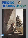
On the Threshold of Illness and Emotional Isolation [PDF - 90 KB - 2 pages]
| EID | Potter P. On the Threshold of Illness and Emotional Isolation. Emerg Infect Dis. 2006;12(5):878-879. https://doi.org/10.3201/eid1205.ac1205 |
|---|---|
| AMA | Potter P. On the Threshold of Illness and Emotional Isolation. Emerging Infectious Diseases. 2006;12(5):878-879. doi:10.3201/eid1205.ac1205. |
| APA | Potter, P. (2006). On the Threshold of Illness and Emotional Isolation. Emerging Infectious Diseases, 12(5), 878-879. https://doi.org/10.3201/eid1205.ac1205. |