Perspective
Clinical Epidemiology of Malaria in the Highlands of Western Kenya
Malaria in the highlands of Kenya is traditionally regarded as unstable and limited by low temperature. Brief warm periods may facilitate malaria transmission and are therefore able to generate epidemic conditions in immunologically naive human populations living at high altitudes. The adult:child ratio (ACR) of malaria admissions is a simple tool we have used to assess the degree of functional immunity in the catchment population of a health facility. Examples of ACR are collected from inpatient admission data at facilities with a range of malaria endemicities in Kenya. Two decades of inpatient malaria admission data from three health facilities in a high-altitude area of western Kenya do not support the canonical view of unstable transmission. The malaria of the region is best described as seasonal and meso-endemic. We discuss the implications for malaria control options in the Kenyan highlands.
| EID | Hay SI, Noor AM, Simba M, Busolo M, Guyatt HL, Ochola SA, et al. Clinical Epidemiology of Malaria in the Highlands of Western Kenya. Emerg Infect Dis. 2002;8(6):543-548. https://doi.org/10.3201/eid0806.010309 |
|---|---|
| AMA | Hay SI, Noor AM, Simba M, et al. Clinical Epidemiology of Malaria in the Highlands of Western Kenya. Emerging Infectious Diseases. 2002;8(6):543-548. doi:10.3201/eid0806.010309. |
| APA | Hay, S. I., Noor, A. M., Simba, M., Busolo, M., Guyatt, H. L., Ochola, S. A....Snow, R. W. (2002). Clinical Epidemiology of Malaria in the Highlands of Western Kenya. Emerging Infectious Diseases, 8(6), 543-548. https://doi.org/10.3201/eid0806.010309. |
Synopses
Parachlamydiaceae: Potential Emerging Pathogens
Parachlamydiaceae, which naturally infect amoebae, form a sister taxon to the Chlamydiaceae on the basis of the Chlamydia-like cycle of replication and 80% to 90% homology of ribosomal RNA genes. Because intra-amoebal growth could increase the virulence of some intracellular bacteria, Parachlamydiaceae may be pathogenic. Arguments supporting a pathogenic role are that Chlamydia pneumoniae, a well-recognized agent of pneumonia, was shown to infect free-living amoebae and that another member of the Chlamydiales, Simkania negevensis, which has 88% homology with Parachlamydia acanthamoebae, has caused pneumonia in adults and acute bronchiolitis in infants. The recent identification of a 16S rRNA gene sequence of a Parachlamydiaceae from bronchoalveolar lavage is additional evidence supporting potential for pathogenicity.
| EID | Greub G, Raoult D. Parachlamydiaceae: Potential Emerging Pathogens. Emerg Infect Dis. 2002;8(6):626-630. https://doi.org/10.3201/eid0806.010210 |
|---|---|
| AMA | Greub G, Raoult D. Parachlamydiaceae: Potential Emerging Pathogens. Emerging Infectious Diseases. 2002;8(6):626-630. doi:10.3201/eid0806.010210. |
| APA | Greub, G., & Raoult, D. (2002). Parachlamydiaceae: Potential Emerging Pathogens. Emerging Infectious Diseases, 8(6), 626-630. https://doi.org/10.3201/eid0806.010210. |
Research
Geographic Association of Rickettsia felis-Infected Opossums with Human Murine Typhus, Texas
Application of molecular diagnostic technology in the past 10 years has resulted in the discovery of several new species of pathogenic rickettsiae, including Rickettsia felis. As more sequence information for rickettsial genes has become available, the data have been used to reclassify rickettsial species and to develop new diagnostic tools for analysis of mixed rickettsial pathogens. R. felis has been associated with opossums and their fleas in Texas and California. Because R. felis can cause human illness, we investigated the distribution dynamics in the murine typhus–endemic areas of these two states. The geographic distribution of R. felis-infected opossum populations in two well-established endemic foci overlaps with that of the reported human cases of murine typhus. Descriptive epidemiologic analysis of 1998 human cases in Corpus Christi, Texas, identified disease patterns consistent with studies done in the 1980s. A close geographic association of seropositive opossums (22% R. felis; 8% R. typhi) with human murine typhus cases was also observed.
| EID | Boostrom A, Beier MS, Macaluso JA, Macaluso KR, Sprenger D, Hayes J, et al. Geographic Association of Rickettsia felis-Infected Opossums with Human Murine Typhus, Texas. Emerg Infect Dis. 2002;8(6):549-554. https://doi.org/10.3201/eid0806.010350 |
|---|---|
| AMA | Boostrom A, Beier MS, Macaluso JA, et al. Geographic Association of Rickettsia felis-Infected Opossums with Human Murine Typhus, Texas. Emerging Infectious Diseases. 2002;8(6):549-554. doi:10.3201/eid0806.010350. |
| APA | Boostrom, A., Beier, M. S., Macaluso, J. A., Macaluso, K. R., Sprenger, D., Hayes, J....Azad, A. F. (2002). Geographic Association of Rickettsia felis-Infected Opossums with Human Murine Typhus, Texas. Emerging Infectious Diseases, 8(6), 549-554. https://doi.org/10.3201/eid0806.010350. |
Defining and Detecting Malaria Epidemics in the Highlands of Western Kenya
Epidemic detection algorithms are being increasingly recommended for malaria surveillance in sub-Saharan Africa. We present the results of applying three simple epidemic detection techniques to routinely collected longitudinal pediatric malaria admissions data from three health facilities in the highlands of western Kenya in the late 1980s and 1990s. The algorithms tested were chosen because they could be feasibly implemented at the health facility level in sub-Saharan Africa. Assumptions of these techniques about the normal distribution of admissions data and the confidence intervals used to define normal years were also investigated. All techniques identified two “epidemic” years in one of the sites. The untransformed Cullen method with standard confidence intervals detected the two “epidemic” years in the remaining two sites but also triggered many false alarms. The performance of these methods is discussed and comments made about their appropriateness for the highlands of western Kenya
| EID | Hay SI, Simba M, Busolo M, Noor AM, Guyatt HL, Ochola SA, et al. Defining and Detecting Malaria Epidemics in the Highlands of Western Kenya. Emerg Infect Dis. 2002;8(6):555-562. https://doi.org/10.3201/eid0806.010310 |
|---|---|
| AMA | Hay SI, Simba M, Busolo M, et al. Defining and Detecting Malaria Epidemics in the Highlands of Western Kenya. Emerging Infectious Diseases. 2002;8(6):555-562. doi:10.3201/eid0806.010310. |
| APA | Hay, S. I., Simba, M., Busolo, M., Noor, A. M., Guyatt, H. L., Ochola, S. A....Snow, R. W. (2002). Defining and Detecting Malaria Epidemics in the Highlands of Western Kenya. Emerging Infectious Diseases, 8(6), 555-562. https://doi.org/10.3201/eid0806.010310. |
Waterborne Outbreak of Norwalk-Like Virus Gastroenteritis at a Tourist Resort, Italy
In July 2000, an outbreak of gastroenteritis occurred at a tourist resort in the Gulf of Taranto in southern Italy. Illness in 344 people, 69 of whom were staff members, met the case definition. Norwalk-like virus (NLV) was found in 22 of 28 stool specimens tested. The source of illness was likely contaminated drinking water, as environmental inspection identified a breakdown in the resort water system and tap water samples were contaminated with fecal bacteria. Attack rates were increased (51.4%) in staff members involved in water sports. Relative risks were significant only for exposure to beach showers and consuming drinks with ice. Although Italy has no surveillance system for nonbacterial gastroenteritis, no outbreak caused by NLV has been described previously in the country.
| EID | Boccia D, Tozzi AE, Cotter B, Rizzo C, Russo T, Buttinelli G, et al. Waterborne Outbreak of Norwalk-Like Virus Gastroenteritis at a Tourist Resort, Italy. Emerg Infect Dis. 2002;8(6):563-568. https://doi.org/10.3201/eid0806.010371 |
|---|---|
| AMA | Boccia D, Tozzi AE, Cotter B, et al. Waterborne Outbreak of Norwalk-Like Virus Gastroenteritis at a Tourist Resort, Italy. Emerging Infectious Diseases. 2002;8(6):563-568. doi:10.3201/eid0806.010371. |
| APA | Boccia, D., Tozzi, A. E., Cotter, B., Rizzo, C., Russo, T., Buttinelli, G....Ruggeri, F. M. (2002). Waterborne Outbreak of Norwalk-Like Virus Gastroenteritis at a Tourist Resort, Italy. Emerging Infectious Diseases, 8(6), 563-568. https://doi.org/10.3201/eid0806.010371. |
Medical Care Capacity for Influenza Outbreaks, Los Angeles
In December 1997, media reported hospital overcrowding and “the worst [flu epidemic] in the past two decades” in Los Angeles County (LAC). We found that rates of pneumonia and influenza deaths, hospitalizations, and claims were substantially higher for the 1997–98 influenza season than the previous six seasons. Hours of emergency medical services (EMS) diversion (when emergency departments could not receive incoming patients) peaked during the influenza seasons studied; the number of EMS diversion hours per season also increased during the seasons 1993–94 to 1997–98, suggesting a decrease in medical care capacity during influenza seasons. Over the seven influenza seasons studied, the number of licensed beds decreased 12%, while the LAC population increased 5%. Our findings suggest that the capacity of health-care systems to handle patient visits during influenza seasons is diminishing.
| EID | Glaser CA, Gilliam S, Thompson WW, Dassey DE, Waterman SH, Saruwatari M, et al. Medical Care Capacity for Influenza Outbreaks, Los Angeles. Emerg Infect Dis. 2002;8(6):569-574. https://doi.org/10.3201/eid0806.010370 |
|---|---|
| AMA | Glaser CA, Gilliam S, Thompson WW, et al. Medical Care Capacity for Influenza Outbreaks, Los Angeles. Emerging Infectious Diseases. 2002;8(6):569-574. doi:10.3201/eid0806.010370. |
| APA | Glaser, C. A., Gilliam, S., Thompson, W. W., Dassey, D. E., Waterman, S. H., Saruwatari, M....Fukuda, K. (2002). Medical Care Capacity for Influenza Outbreaks, Los Angeles. Emerging Infectious Diseases, 8(6), 569-574. https://doi.org/10.3201/eid0806.010370. |
Drought-Induced Amplification of Saint Louis encephalitis virus, Florida
We used a dynamic hydrology model to simulate water table depth (WTD) and quantify the relationship between Saint Louis encephalitis virus (SLEV) transmission and hydrologic conditions in Indian River County, Florida, from 1986 through 1991, a period with an SLEV epidemic. Virus transmission followed periods of modeled drought (specifically low WTDs 12 to 17 weeks before virus transmission, followed by a rising of the water table 1 to 2 weeks before virus transmission). Further evidence from collections of Culex nigripalpus (the major mosquito vector of SLEV in Florida) suggests that during extended spring droughts vector mosquitoes and nestling, juvenile, and adult wild birds congregate in selected refuges, facilitating epizootic amplification of SLEV. When the drought ends and habitat availability increases, the SLEV-infected Cx. nigripalpus and wild birds disperse, initiating an SLEV transmission cycle. These findings demonstrate a mechanism by which drought facilitates the amplification of SLEV and its subsequent transmission to humans.
| EID | Shaman J, Day JF, Stieglitz M. Drought-Induced Amplification of Saint Louis encephalitis virus, Florida. Emerg Infect Dis. 2002;8(6):575-580. https://doi.org/10.3201/eid0806.010417 |
|---|---|
| AMA | Shaman J, Day JF, Stieglitz M. Drought-Induced Amplification of Saint Louis encephalitis virus, Florida. Emerging Infectious Diseases. 2002;8(6):575-580. doi:10.3201/eid0806.010417. |
| APA | Shaman, J., Day, J. F., & Stieglitz, M. (2002). Drought-Induced Amplification of Saint Louis encephalitis virus, Florida. Emerging Infectious Diseases, 8(6), 575-580. https://doi.org/10.3201/eid0806.010417. |
Epidemiologic Differences Between Cyclosporiasis and Cryptosporidiosis in Peruvian Children
We compared the epidemiologic characteristics of cyclosporiasis and cryptosporidiosis in data from a cohort study of diarrhea in a periurban community near Lima, Peru. Children had an average of 0.20 episodes of cyclosporiasis/year and 0.22 episodes of cryptosporidiosis/year of follow-up. The incidence of cryptosporidiosis peaked at 0.42 for 1-year-old children and declined to 0.06 episodes/child-year for 5- to 9-year-old children. In contrast, the incidence of cyclosporiasis was fairly constant among 1- to 9-year-old children (0.21 to 0.28 episodes/child-year). Likelihood of diarrhea decreased significantly with each episode of cyclosporiasis; for cryptosporidiosis, this trend was not statistically significant. Both infections were more frequent during the warm season (December to May) than the cooler season (June to November). Cryptosporidiosis was more frequent in children from houses without a latrine or toilet. Cyclosporiasis was associated with ownership of domestic animals, especially birds, guinea pigs, and rabbits.
| EID | Bern C, Ortega Y, Checkley W, Roberts JM, Lescano AG, Cabrera L, et al. Epidemiologic Differences Between Cyclosporiasis and Cryptosporidiosis in Peruvian Children. Emerg Infect Dis. 2002;8(6):581-585. https://doi.org/10.3201/eid0806.010331 |
|---|---|
| AMA | Bern C, Ortega Y, Checkley W, et al. Epidemiologic Differences Between Cyclosporiasis and Cryptosporidiosis in Peruvian Children. Emerging Infectious Diseases. 2002;8(6):581-585. doi:10.3201/eid0806.010331. |
| APA | Bern, C., Ortega, Y., Checkley, W., Roberts, J. M., Lescano, A. G., Cabrera, L....Gilman, R. H. (2002). Epidemiologic Differences Between Cyclosporiasis and Cryptosporidiosis in Peruvian Children. Emerging Infectious Diseases, 8(6), 581-585. https://doi.org/10.3201/eid0806.010331. |
Population-Based Study of Acute Respiratory Infections in Children, Greenland
Acute respiratory infections (ARI) are frequent in Inuit children, in terms of incidence and severity. A cohort of 294 children <2 years of age was formed in Sisimiut, a community on the west coast of Greenland, and followed from 1996 to 1998. Data on ARI were collected during weekly visits at home and child-care centers; visits to the community health center were also recorded. The cohort had respiratory symptoms on 41.6% and fever on 4.9% of surveyed days. The incidence of upper and lower respiratory tract infections was 1.6 episodes and 0.9 episodes per 100 days at risk, respectively. Up to 65% of the episodes of ARI caused activity restriction; 40% led to contact with the health center. Compared with studies from other parts of the world, the incidence of ARI appears to be high in Inuit children.
| EID | Koch A, Sørensen P, Homøe P, Mølbak K, Pedersen FK, Mortensen T, et al. Population-Based Study of Acute Respiratory Infections in Children, Greenland. Emerg Infect Dis. 2002;8(6):586-593. https://doi.org/10.3201/eid0806.010321 |
|---|---|
| AMA | Koch A, Sørensen P, Homøe P, et al. Population-Based Study of Acute Respiratory Infections in Children, Greenland. Emerging Infectious Diseases. 2002;8(6):586-593. doi:10.3201/eid0806.010321. |
| APA | Koch, A., Sørensen, P., Homøe, P., Mølbak, K., Pedersen, F. K., Mortensen, T....Melbye, M. (2002). Population-Based Study of Acute Respiratory Infections in Children, Greenland. Emerging Infectious Diseases, 8(6), 586-593. https://doi.org/10.3201/eid0806.010321. |
Streptococcus pneumoniae, Brooklyn, New York: Fluoroquinolone Resistance at our Doorstep
To examine the resistance rates and epidemiology of Streptococcus pneumoniae in Brooklyn, New York, isolates were collected during two boroughwide surveillance periods in 1997 and 1999. Of 138 isolates, 67% were susceptible to penicillin and 34% to ciprofloxacin. Susceptibility rates to ciprofloxacin decreased dramatically from 1997 to 1999 (47% to 16%, p=0.0003). Five isolates (3.6%) were resistant to levofloxacin. Western Brooklyn had lower rates of susceptibility to penicillin compared with eastern neighborhoods. More isolates in the eastern neighborhoods belonged to the Spanish/French 9/14 clone, and isolates in the western neighborhoods tended to belong to the Spanish/USA 23F clone. Residents of the western neighborhoods were more likely to be white and elderly and less likely to be receiving Medicaid or public assistance, characteristics associated with increased health-care and antibiotic use. Brooklyn residents appear to be at high risk for fluoroquinolone-resistant S. pneumoniae. Our results underscore the need for vigilant regional surveillance.
| EID | Quale J, Landman D, Ravishankar J, Flores C, Bratu S. Streptococcus pneumoniae, Brooklyn, New York: Fluoroquinolone Resistance at our Doorstep. Emerg Infect Dis. 2002;8(6):594-597. https://doi.org/10.3201/eid0806.010275 |
|---|---|
| AMA | Quale J, Landman D, Ravishankar J, et al. Streptococcus pneumoniae, Brooklyn, New York: Fluoroquinolone Resistance at our Doorstep. Emerging Infectious Diseases. 2002;8(6):594-597. doi:10.3201/eid0806.010275. |
| APA | Quale, J., Landman, D., Ravishankar, J., Flores, C., & Bratu, S. (2002). Streptococcus pneumoniae, Brooklyn, New York: Fluoroquinolone Resistance at our Doorstep. Emerging Infectious Diseases, 8(6), 594-597. https://doi.org/10.3201/eid0806.010275. |
Mycobacterium tuberculosis: An Emerging Disease of Free-Ranging Wildlife
Expansion of ecotourism-based industries, changes in land-use practices, and escalating competition for resources have increased contact between free-ranging wildlife and humans. Although human presence in wildlife areas may provide an important economic benefit through ecotourism, exposure to human pathogens may represent a health risk for wildlife. This report is the first to document introduction of a primary human pathogen into free-ranging wildlife. We describe outbreaks of Mycobacterium tuberculosis, a human pathogen, in free-ranging banded mongooses (Mungos mungo) in Botswana and suricates (Suricata suricatta) in South Africa. Wildlife managers and scientists must address the potential threat that humans pose to the health of free-ranging wildlife.
| EID | Alexander KA, Pleydell E, Williams MC, Lane EP, Nyange JF, Michel AL. Mycobacterium tuberculosis: An Emerging Disease of Free-Ranging Wildlife. Emerg Infect Dis. 2002;8(6):598-601. https://doi.org/10.3201/eid0806.010358 |
|---|---|
| AMA | Alexander KA, Pleydell E, Williams MC, et al. Mycobacterium tuberculosis: An Emerging Disease of Free-Ranging Wildlife. Emerging Infectious Diseases. 2002;8(6):598-601. doi:10.3201/eid0806.010358. |
| APA | Alexander, K. A., Pleydell, E., Williams, M. C., Lane, E. P., Nyange, J. F., & Michel, A. L. (2002). Mycobacterium tuberculosis: An Emerging Disease of Free-Ranging Wildlife. Emerging Infectious Diseases, 8(6), 598-601. https://doi.org/10.3201/eid0806.010358. |
Community-Acquired Methicillin-Resistant Staphylococcus aureus, Finland
Methicillin-resistant Staphylococcus aureus (MRSA) is no longer only hospital acquired. MRSA is defined as community acquired if the MRSA-positive specimen was obtained outside hospital settings or within 2 days of hospital admission, and if it was from a person who had not been hospitalized within 2 years before the date of MRSA isolation. To estimate the proportion of community-acquired MRSA, we analyzed previous hospitalizations for all MRSA-positive persons in Finland from1997 to 1999 by using data from the National Hospital Discharge Register. Of 526 MRSA-positive persons, 21% had community-acquired MRSA. Three MRSA strains identified by phage typing, pulsed-field gel electrophoresis, and ribotyping were associated with community acquisition. None of the strains were multiresistant, and all showed an mec hypervariable region hybridization pattern A (HVR type A). None of the epidemic multiresistant hospital strains were prevalent in nonhospitalized persons. Our population-based data suggest that community-acquired MRSA may also arise de novo, through horizontal acquisition of the mecA gene.
| EID | Salmenlinna S, Lyytikäinen O, Vuopio-Varkila J. Community-Acquired Methicillin-Resistant Staphylococcus aureus, Finland. Emerg Infect Dis. 2002;8(6):602-607. https://doi.org/10.3201/eid0806.010313 |
|---|---|
| AMA | Salmenlinna S, Lyytikäinen O, Vuopio-Varkila J. Community-Acquired Methicillin-Resistant Staphylococcus aureus, Finland. Emerging Infectious Diseases. 2002;8(6):602-607. doi:10.3201/eid0806.010313. |
| APA | Salmenlinna, S., Lyytikäinen, O., & Vuopio-Varkila, J. (2002). Community-Acquired Methicillin-Resistant Staphylococcus aureus, Finland. Emerging Infectious Diseases, 8(6), 602-607. https://doi.org/10.3201/eid0806.010313. |
Neurocysticercosis in Radiographically Imaged Seizure Patients in U.S. Emergency Departments
Neurocysticercosis appears to be on the rise in the United States, based on immigration patterns and published cases series, including reports of domestic acquisition. We used a collaborative network of U.S. emergency departments to characterize the epidemiology of neurocysticercosis in seizure patients. Data were collected prospectively at 11 university-affiliated, geographically diverse, urban U.S. emergency departments from July 1996 to September 1998. Patients with a seizure who underwent neuroimaging were included. Of the 1,801 patients enrolled in the study, 38 (2.1%) had seizures attributable to neurocysticercosis. The disease was detected in 9 of the 11 sites and was associated with Hispanic ethnicity, immigrant status, and exposure to areas where neurocysticercosis is endemic. This disease appears to be widely distributed and highly prevalent in certain populations (e.g., Hispanic patients) and areas (e.g., Southwest).
| EID | Ong S, Talan DA, Moran GJ, Mower WR, Newdow M, Tsang VC, et al. Neurocysticercosis in Radiographically Imaged Seizure Patients in U.S. Emergency Departments. Emerg Infect Dis. 2002;8(6):608-613. https://doi.org/10.3201/eid0806.010377 |
|---|---|
| AMA | Ong S, Talan DA, Moran GJ, et al. Neurocysticercosis in Radiographically Imaged Seizure Patients in U.S. Emergency Departments. Emerging Infectious Diseases. 2002;8(6):608-613. doi:10.3201/eid0806.010377. |
| APA | Ong, S., Talan, D. A., Moran, G. J., Mower, W. R., Newdow, M., Tsang, V. C....Pinner, R. W. (2002). Neurocysticercosis in Radiographically Imaged Seizure Patients in U.S. Emergency Departments. Emerging Infectious Diseases, 8(6), 608-613. https://doi.org/10.3201/eid0806.010377. |
Two New Rhabdoviruses (Rhabdoviridae) Isolated from Birds During Surveillance for Arboviral Encephalitis, Northeastern United States
Two novel rhabdoviruses were isolated from birds during surveillance for arboviral encephalitis in the northeastern United States. The first, designated Farmington virus, is a tentative new member of the Vesiculovirus genus. The second, designated Rhode Island virus, is unclassified antigenically, but its ultrastructure and size are more similar to those of some of the plant rhabdoviruses. Both viruses infect birds and mice, as well as monkey kidney cells in culture, but their importance for human health is unknown.
| EID | Travassos da Rosa AP, Mather TN, Takeda T, Whitehouse CA, Shope RE, Popov VL, et al. Two New Rhabdoviruses (Rhabdoviridae) Isolated from Birds During Surveillance for Arboviral Encephalitis, Northeastern United States. Emerg Infect Dis. 2002;8(6):616-618. https://doi.org/10.3201/eid0806.010384 |
|---|---|
| AMA | Travassos da Rosa AP, Mather TN, Takeda T, et al. Two New Rhabdoviruses (Rhabdoviridae) Isolated from Birds During Surveillance for Arboviral Encephalitis, Northeastern United States. Emerging Infectious Diseases. 2002;8(6):616-618. doi:10.3201/eid0806.010384. |
| APA | Travassos da Rosa, A. P., Mather, T. N., Takeda, T., Whitehouse, C. A., Shope, R. E., Popov, V. L....Tesh, R. B. (2002). Two New Rhabdoviruses (Rhabdoviridae) Isolated from Birds During Surveillance for Arboviral Encephalitis, Northeastern United States. Emerging Infectious Diseases, 8(6), 616-618. https://doi.org/10.3201/eid0806.010384. |
Cryptosporidium Oocysts in a Water Supply Associated with a Cryptosporidiosis Outbreak
An outbreak of cryptosporidiosis occurred in and around Clitheroe, Lancashire, in northwest England, during March 2000. Fifty-eight cases of diarrhea with Cryptosporidium identified in stool specimens were reported. Cryptosporidium oocysts were identified in samples from the water treatment works as well as domestic taps. Descriptive epidemiology suggested that drinking unboiled tap water in a single water zone was the common factor linking cases. Environmental investigation suggested that contamination with animal feces was the likely source of the outbreak. This outbreak was unusual in that hydrodynamic modeling was used to give a good estimate of the peak oocyst count at the time of the contamination incident. The oocysts’ persistence in the water distribution system after switching to another water source was also unusual. This persistence may have been due to oocysts being entrapped within biofilm. Despite the continued presence of oocysts, epidemiologic evidence suggested that no one became ill after the water source was changed.
| EID | Howe AD, Forster S, Morton S, Marshall R, Osborn KS, Wright P, et al. Cryptosporidium Oocysts in a Water Supply Associated with a Cryptosporidiosis Outbreak. Emerg Infect Dis. 2002;8(6):619-624. https://doi.org/10.3201/eid0806.010271 |
|---|---|
| AMA | Howe AD, Forster S, Morton S, et al. Cryptosporidium Oocysts in a Water Supply Associated with a Cryptosporidiosis Outbreak. Emerging Infectious Diseases. 2002;8(6):619-624. doi:10.3201/eid0806.010271. |
| APA | Howe, A. D., Forster, S., Morton, S., Marshall, R., Osborn, K. S., Wright, P....Hunter, P. R. (2002). Cryptosporidium Oocysts in a Water Supply Associated with a Cryptosporidiosis Outbreak. Emerging Infectious Diseases, 8(6), 619-624. https://doi.org/10.3201/eid0806.010271. |
Dispatches
Three Drinking-Water–Associated Cryptosporidiosis Outbreaks, Northern Ireland
Three recent drinking-water–associated cryptosporidiosis outbreaks in Northern Ireland were investigated by using genotyping and subgenotyping tools. One Cryptosporidium parvum outbreak was caused by the bovine genotype, and two were caused by the human genotype. Subgenotyping analyses indicate that two predominant subgenotypes were associated with these outbreaks and had been circulating in the community.
| EID | Glaberman S, Moore JE, Lowery CJ, Chalmers RM, Sulaiman I, Elwin K, et al. Three Drinking-Water–Associated Cryptosporidiosis Outbreaks, Northern Ireland. Emerg Infect Dis. 2002;8(6):631-633. https://doi.org/10.3201/eid0806.010368 |
|---|---|
| AMA | Glaberman S, Moore JE, Lowery CJ, et al. Three Drinking-Water–Associated Cryptosporidiosis Outbreaks, Northern Ireland. Emerging Infectious Diseases. 2002;8(6):631-633. doi:10.3201/eid0806.010368. |
| APA | Glaberman, S., Moore, J. E., Lowery, C. J., Chalmers, R. M., Sulaiman, I., Elwin, K....Xiao, L. (2002). Three Drinking-Water–Associated Cryptosporidiosis Outbreaks, Northern Ireland. Emerging Infectious Diseases, 8(6), 631-633. https://doi.org/10.3201/eid0806.010368. |
Cluster of African Trypanosomiasis in Travelers to Tanzanian National Parks
Game parks in Tanzania have long been considered to be at low risk for African trypanosomiasis; however, nine cases of the disease associated with these parks were recently reported. The outbreak was detected through TropNetEurop, a sentinel surveillance network of clinical sites throughout Europe.
| EID | Jelinek T, Bisoffi Z, Bonazzi L, van Thiel P, Bronner U, de Frey A, et al. Cluster of African Trypanosomiasis in Travelers to Tanzanian National Parks. Emerg Infect Dis. 2002;8(6):634-635. https://doi.org/10.3201/eid0806.010432 |
|---|---|
| AMA | Jelinek T, Bisoffi Z, Bonazzi L, et al. Cluster of African Trypanosomiasis in Travelers to Tanzanian National Parks. Emerging Infectious Diseases. 2002;8(6):634-635. doi:10.3201/eid0806.010432. |
| APA | Jelinek, T., Bisoffi, Z., Bonazzi, L., van Thiel, P., Bronner, U., de Frey, A....Ripamont, D. (2002). Cluster of African Trypanosomiasis in Travelers to Tanzanian National Parks. Emerging Infectious Diseases, 8(6), 634-635. https://doi.org/10.3201/eid0806.010432. |
Excretion of Vancomycin-Resistant Enterococci by Wild Mammals
A survey of fecal samples found enterococcal excretion in 82% of 388 bank voles (Clethrionomys glareolus), 92% of 131 woodmice (Apodemus sylvaticus), and 75% of 165 badgers (Meles meles). Vancomycin-resistant enterococci, all Enterococcus faecium of vanA genotype, were excreted by 4.6% of the woodmice and 1.2% of the badgers, but by none of the bank voles.
| EID | Mallon D, Corkill JE, Hazel SM, Wilson J, French NP, Bennett M, et al. Excretion of Vancomycin-Resistant Enterococci by Wild Mammals. Emerg Infect Dis. 2002;8(6):636-638. https://doi.org/10.3201/eid0806.010247 |
|---|---|
| AMA | Mallon D, Corkill JE, Hazel SM, et al. Excretion of Vancomycin-Resistant Enterococci by Wild Mammals. Emerging Infectious Diseases. 2002;8(6):636-638. doi:10.3201/eid0806.010247. |
| APA | Mallon, D., Corkill, J. E., Hazel, S. M., Wilson, J., French, N. P., Bennett, M....Hart, C. (2002). Excretion of Vancomycin-Resistant Enterococci by Wild Mammals. Emerging Infectious Diseases, 8(6), 636-638. https://doi.org/10.3201/eid0806.010247. |
Fatal Infection of a Pet Monkey with Human herpesvirus 1
Concerns have been raised about pet monkeys as a potential threat to humans. We report the opposite situation, a danger to pets that arises from humans. Similar to herpesvirus B (Cercopithecine herpesvirus 1), which endangers humans but not its host species, Human herpesvirus 1 can act as a “killer virus” when crossing the species barrier to New World monkeys.
| EID | Huemer HP, Larcher C, Czedik-Eysenberg T, Nowotny N, Reifinger M. Fatal Infection of a Pet Monkey with Human herpesvirus 1. Emerg Infect Dis. 2002;8(6):639-641. https://doi.org/10.3201/eid0806.010341 |
|---|---|
| AMA | Huemer HP, Larcher C, Czedik-Eysenberg T, et al. Fatal Infection of a Pet Monkey with Human herpesvirus 1. Emerging Infectious Diseases. 2002;8(6):639-641. doi:10.3201/eid0806.010341. |
| APA | Huemer, H. P., Larcher, C., Czedik-Eysenberg, T., Nowotny, N., & Reifinger, M. (2002). Fatal Infection of a Pet Monkey with Human herpesvirus 1. Emerging Infectious Diseases, 8(6), 639-641. https://doi.org/10.3201/eid0806.010341. |
Letters
Serologic Evidence of Human Granulocytic Ehrlichiosis, Greece
| EID | Daniel SA, Manika K, Arvanitidou M, Diza E, Symeonidis N, Antoniadis A. Serologic Evidence of Human Granulocytic Ehrlichiosis, Greece. Emerg Infect Dis. 2002;8(6):643-644. https://doi.org/10.3201/eid0806.010500 |
|---|---|
| AMA | Daniel SA, Manika K, Arvanitidou M, et al. Serologic Evidence of Human Granulocytic Ehrlichiosis, Greece. Emerging Infectious Diseases. 2002;8(6):643-644. doi:10.3201/eid0806.010500. |
| APA | Daniel, S. A., Manika, K., Arvanitidou, M., Diza, E., Symeonidis, N., & Antoniadis, A. (2002). Serologic Evidence of Human Granulocytic Ehrlichiosis, Greece. Emerging Infectious Diseases, 8(6), 643-644. https://doi.org/10.3201/eid0806.010500. |
Hantavirus Infection with Marked Sinus Bradycardia, Taiwan
| EID | Liu Y, Huang J, Hsueh P, Luh K. Hantavirus Infection with Marked Sinus Bradycardia, Taiwan. Emerg Infect Dis. 2002;8(6):644-645. https://doi.org/10.3201/eid0806.010340 |
|---|---|
| AMA | Liu Y, Huang J, Hsueh P, et al. Hantavirus Infection with Marked Sinus Bradycardia, Taiwan. Emerging Infectious Diseases. 2002;8(6):644-645. doi:10.3201/eid0806.010340. |
| APA | Liu, Y., Huang, J., Hsueh, P., & Luh, K. (2002). Hantavirus Infection with Marked Sinus Bradycardia, Taiwan. Emerging Infectious Diseases, 8(6), 644-645. https://doi.org/10.3201/eid0806.010340. |
Books and Media
A Dictionary of Virology, Third Edition
| EID | Calisher CH. A Dictionary of Virology, Third Edition. Emerg Infect Dis. 2002;8(6):646. https://doi.org/10.3201/eid0806.020058 |
|---|---|
| AMA | Calisher CH. A Dictionary of Virology, Third Edition. Emerging Infectious Diseases. 2002;8(6):646. doi:10.3201/eid0806.020058. |
| APA | Calisher, C. H. (2002). A Dictionary of Virology, Third Edition. Emerging Infectious Diseases, 8(6), 646. https://doi.org/10.3201/eid0806.020058. |
Corrections
Correction Vol. 8, No. 3
| EID | Correction Vol. 8, No. 3. Emerg Infect Dis. 2002;8(6):554. https://doi.org/10.3201/eid0806.c10806 |
|---|---|
| AMA | Correction Vol. 8, No. 3. Emerging Infectious Diseases. 2002;8(6):554. doi:10.3201/eid0806.c10806. |
| APA | (2002). Correction Vol. 8, No. 3. Emerging Infectious Diseases, 8(6), 554. https://doi.org/10.3201/eid0806.c10806. |
About the Cover
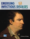
Samuel Johnson (circa 1769)
| EID | Potter P. Samuel Johnson (circa 1769). Emerg Infect Dis. 2002;8(6):648. https://doi.org/10.3201/eid0806.ac0806 |
|---|---|
| AMA | Potter P. Samuel Johnson (circa 1769). Emerging Infectious Diseases. 2002;8(6):648. doi:10.3201/eid0806.ac0806. |
| APA | Potter, P. (2002). Samuel Johnson (circa 1769). Emerging Infectious Diseases, 8(6), 648. https://doi.org/10.3201/eid0806.ac0806. |


