Perspective
Passive Antibody Administration (Immediate Immunity) as a Specific Defense Against Biological Weapons [PDF - 288 KB - 9 pages]
The potential threat of biological warfare with a specific agent is proportional to the susceptibility of the population to that agent. Preventing disease after exposure to a biological agent is partially a function of the immunity of the exposed individual. The only available countermeasure that can provide immediate immunity against a biological agent is passive antibody. Unlike vaccines, which require time to induce protective immunity and depend on the host’s ability to mount an immune response, passive antibody can theoretically confer protection regardless of the immune status of the host. Passive antibody therapy has substantial advantages over antimicrobial agents and other measures for postexposure prophylaxis, including low toxicity and high specific activity. Specific antibodies are active against the major agents of bioterrorism, including anthrax, smallpox, botulinum toxin, tularemia, and plague. This article proposes a biological defense initiative based on developing, producing, and stockpiling specific antibody reagents that can be used to protect the population against biological warfare threats.
| EID | Casadevall A. Passive Antibody Administration (Immediate Immunity) as a Specific Defense Against Biological Weapons. Emerg Infect Dis. 2002;8(8):833-841. https://doi.org/10.3201/eid0808.010516 |
|---|---|
| AMA | Casadevall A. Passive Antibody Administration (Immediate Immunity) as a Specific Defense Against Biological Weapons. Emerging Infectious Diseases. 2002;8(8):833-841. doi:10.3201/eid0808.010516. |
| APA | Casadevall, A. (2002). Passive Antibody Administration (Immediate Immunity) as a Specific Defense Against Biological Weapons. Emerging Infectious Diseases, 8(8), 833-841. https://doi.org/10.3201/eid0808.010516. |
Synopses
Worldwide Occurrence of Beijing/W Strains of Mycobacterium tuberculosis: A Systematic Review [PDF - 298 KB - 7 pages]
Strains of the Beijing/W genotype family of Mycobacterium tuberculosis have caused large outbreaks of tuberculosis, sometimes involving multidrug resistance. This genetically highly conserved family of M. tuberculosis strains predominates in some geographic areas. We have conducted a systematic review of the published reports on these strains to determine their worldwide distribution, spread, and association with drug resistance. Sixteen studies reported prevalence of Beijing strains defined by spoligotyping; another 10 used other definitions. Beijing strains were most prevalent in Asia but were found worldwide. Associations with drug resistance varied: in New York, Cuba, Estonia, and Vietnam, Beijing strains were strongly associated with drug resistance, but elsewhere the association was weak or absent. Although few reports have measured trends in prevalence, the ubiquity of the Beijing strains and their frequent association with outbreaks and drug resistance underline their importance.
| EID | Glynn JR, Whiteley J, Bifani PJ, Kremer K, van Soolingen D. Worldwide Occurrence of Beijing/W Strains of Mycobacterium tuberculosis: A Systematic Review. Emerg Infect Dis. 2002;8(8):843-849. https://doi.org/10.3201/eid0808.020002 |
|---|---|
| AMA | Glynn JR, Whiteley J, Bifani PJ, et al. Worldwide Occurrence of Beijing/W Strains of Mycobacterium tuberculosis: A Systematic Review. Emerging Infectious Diseases. 2002;8(8):843-849. doi:10.3201/eid0808.020002. |
| APA | Glynn, J. R., Whiteley, J., Bifani, P. J., Kremer, K., & van Soolingen, D. (2002). Worldwide Occurrence of Beijing/W Strains of Mycobacterium tuberculosis: A Systematic Review. Emerging Infectious Diseases, 8(8), 843-849. https://doi.org/10.3201/eid0808.020002. |
Research
Use of Automated Ambulatory-Care Encounter Records for Detection of Acute Illness Clusters, Including Potential Bioterrorism Events [PDF - 383 KB - 8 pages]
The advent of domestic bioterrorism has emphasized the need for enhanced detection of clusters of acute illness. We describe a monitoring system operational in eastern Massachusetts, based on diagnoses obtained from electronic records of ambulatory-care encounters. Within 24 hours, ambulatory and telephone encounters recording patients with diagnoses of interest are identified and merged into major syndrome groups. Counts of new episodes of illness, rates calculated from health insurance records, and estimates of the probability of observing at least this number of new episodes are reported for syndrome surveillance. Census tracts with unusually large counts are identified by comparing observed with expected syndrome frequencies. During 1996–1999, weekly counts of new cases of lower respiratory syndrome were highly correlated with weekly hospital admissions. This system complements emergency room- and hospital-based surveillance by adding the capacity to rapidly identify clusters of illness, including potential bioterrorism events.
| EID | Lazarus R, Kleinman K, Dashevsky I, Adams C, Kludt P, DeMaria A, et al. Use of Automated Ambulatory-Care Encounter Records for Detection of Acute Illness Clusters, Including Potential Bioterrorism Events. Emerg Infect Dis. 2002;8(8):753-760. https://doi.org/10.3201/eid0808.020239 |
|---|---|
| AMA | Lazarus R, Kleinman K, Dashevsky I, et al. Use of Automated Ambulatory-Care Encounter Records for Detection of Acute Illness Clusters, Including Potential Bioterrorism Events. Emerging Infectious Diseases. 2002;8(8):753-760. doi:10.3201/eid0808.020239. |
| APA | Lazarus, R., Kleinman, K., Dashevsky, I., Adams, C., Kludt, P., DeMaria, A....Platt, R. (2002). Use of Automated Ambulatory-Care Encounter Records for Detection of Acute Illness Clusters, Including Potential Bioterrorism Events. Emerging Infectious Diseases, 8(8), 753-760. https://doi.org/10.3201/eid0808.020239. |
Outbreak of Serogroup W135 Meningococcal Disease after the Hajj Pilgrimage, Europe, 2000 [PDF - 1.18 MB - 8 pages]
The 2000 Hajj (March 15–18) was followed by an outbreak of Neisseria meningitidis W135 2a: P1.2,5 in Europe. From March 18 to July 31, 2000, some 90 cases of meningococcal infection were reported from nine countries, mostly the United Kingdom (UK) and France; 14 cases were fatal. Although most early cases were in pilgrims, the outbreak spread to their contacts and then to those with no known pilgrim contact. In France and the UK, the outbreak case-fatality rate was compared with the rate reported from national surveillance. The risk of dying during this outbreak was higher in France and the UK, although the difference was not statistically significant. Prophylaxis for all pilgrims and their household contacts was offered in France; in the UK and other European countries, prophylaxis was recommended only for close contacts. No difference in transmission rates following intervention was detected between France and the UK.
| EID | Aguilera J, Perrocheau A, Meffre C, Hahné S. Outbreak of Serogroup W135 Meningococcal Disease after the Hajj Pilgrimage, Europe, 2000. Emerg Infect Dis. 2002;8(8):761-767. https://doi.org/10.3201/eid0808.010422 |
|---|---|
| AMA | Aguilera J, Perrocheau A, Meffre C, et al. Outbreak of Serogroup W135 Meningococcal Disease after the Hajj Pilgrimage, Europe, 2000. Emerging Infectious Diseases. 2002;8(8):761-767. doi:10.3201/eid0808.010422. |
| APA | Aguilera, J., Perrocheau, A., Meffre, C., & Hahné, S. (2002). Outbreak of Serogroup W135 Meningococcal Disease after the Hajj Pilgrimage, Europe, 2000. Emerging Infectious Diseases, 8(8), 761-767. https://doi.org/10.3201/eid0808.010422. |
Genetic Characterization of Hantaviruses Transmitted by the Korean Field Mouse (Apodemus peninsulae), Far East Russia [PDF - 381 KB - 9 pages]
In an epizootiologic survey of 122 rodents captured in Vladivostok, Russia, antibodies positive for hantavirus were found in Apodemus peninsulae (4/70), A. agrarius (1/39), and Clethrionomys rufocanus (1/8). The hantavirus sequences identified in two seropositive A. peninsulae and two patients with hemorrhagic fever with renal syndrome (HFRS) from the Primorye region of Far East Russia were designated as Solovey and Primorye, respectively. The nucleotide sequences of the Solovey, Primorye, and Amur (obtained through GenBank) sequences were closely related (>92% identity). Solovey and Primorye sequences shared 84% nucleotide identity with the prototype Hantaan 76-118. Phylogenetic analysis also indicated a close relationship between Solovey, Primorye, Amur, and other viruses identified in Russia, China, and Korea. Our findings suggest that the Korean field mouse (A. peninsulae) is the reservoir for a hantavirus that causes HFRS over a vast area of east Asia, including Far East Russia.
| EID | Lokugamage K, Kariwa H, Hayasaka D, Cui BZ, Iwasaki T, Lokugamage N, et al. Genetic Characterization of Hantaviruses Transmitted by the Korean Field Mouse (Apodemus peninsulae), Far East Russia. Emerg Infect Dis. 2002;8(8):768-776. https://doi.org/10.3201/eid0808.010494 |
|---|---|
| AMA | Lokugamage K, Kariwa H, Hayasaka D, et al. Genetic Characterization of Hantaviruses Transmitted by the Korean Field Mouse (Apodemus peninsulae), Far East Russia. Emerging Infectious Diseases. 2002;8(8):768-776. doi:10.3201/eid0808.010494. |
| APA | Lokugamage, K., Kariwa, H., Hayasaka, D., Cui, B. Z., Iwasaki, T., Lokugamage, N....Takashima, I. (2002). Genetic Characterization of Hantaviruses Transmitted by the Korean Field Mouse (Apodemus peninsulae), Far East Russia. Emerging Infectious Diseases, 8(8), 768-776. https://doi.org/10.3201/eid0808.010494. |
West Nile Virus Outbreak in Horses, Southern France, 2000: Results of a Serosurvey
During late summer and autumn 2000, a West Nile fever outbreak in southern France resulted in 76 equine clinical cases; 21 horses died. We report the results of a large serosurvey of all equines within a 10-km radius of laboratory-confirmed cases. Blood samples were obtained from 5,107 equines, distributed in groups of 1 to 91 animals. West Nile virus immunoglobulin (Ig) G antibodies were found in 8.5% of animals (n=432). Forty-two percent of the IgG-positive animals were also IgM positive. Horses living in small groups were more affected than those in large groups. The results suggest that West Nile virus is not endemic in the affected area, the Camargue; rather, sporadic outbreaks are separated by long silent periods.
| EID | Durand B, Chevalier V, Pouillot R, Labie J, Marendat I, Murgue B, et al. West Nile Virus Outbreak in Horses, Southern France, 2000: Results of a Serosurvey. Emerg Infect Dis. 2002;8(8):777-782. https://doi.org/10.3201/eid0808.010486 |
|---|---|
| AMA | Durand B, Chevalier V, Pouillot R, et al. West Nile Virus Outbreak in Horses, Southern France, 2000: Results of a Serosurvey. Emerging Infectious Diseases. 2002;8(8):777-782. doi:10.3201/eid0808.010486. |
| APA | Durand, B., Chevalier, V., Pouillot, R., Labie, J., Marendat, I., Murgue, B....Zientara, S. (2002). West Nile Virus Outbreak in Horses, Southern France, 2000: Results of a Serosurvey. Emerging Infectious Diseases, 8(8), 777-782. https://doi.org/10.3201/eid0808.010486. |
Outbreak of Cyclosporiasis Associated with Imported Raspberries, Philadelphia, Pennsylvania, 2000 [PDF - 510 KB - 6 pages]
An outbreak of cyclosporiasis occurred in attendees of a wedding reception held in Philadelphia, Pennsylvania, on June 10, 2000. In a retrospective cohort study, 54 (68.4%) of the 79 interviewed guests and members of the wedding party met the case definition. The wedding cake, which had a cream filling that included raspberries, was the food item most strongly associated with illness (multivariate relative risk, 5.9; 95% confidence interval, 3.6 to 10.5). Leftover cake was positive for Cyclospora DNA by polymerase chain reaction analyses. Sequencing of the amplified fragments confirmed that the organism was Cyclospora cayetanensis. The year 2000 was the fifth year since 1995 that outbreaks of cyclosporiasis definitely or probably associated with Guatemalan raspberries have occurred in the spring in North America. Additionally, this is the second documented U.S. outbreak, and the first associated with raspberries, for which Cyclospora has been detected in the epidemiologically implicated food item.
| EID | Ho AY, Lopez AS, Eberhart MG, Levenson R, Finkel BS, da Silva AJ, et al. Outbreak of Cyclosporiasis Associated with Imported Raspberries, Philadelphia, Pennsylvania, 2000. Emerg Infect Dis. 2002;8(8):783-788. https://doi.org/10.3201/eid0808.020012 |
|---|---|
| AMA | Ho AY, Lopez AS, Eberhart MG, et al. Outbreak of Cyclosporiasis Associated with Imported Raspberries, Philadelphia, Pennsylvania, 2000. Emerging Infectious Diseases. 2002;8(8):783-788. doi:10.3201/eid0808.020012. |
| APA | Ho, A. Y., Lopez, A. S., Eberhart, M. G., Levenson, R., Finkel, B. S., da Silva, A. J....Herwaldt, B. L. (2002). Outbreak of Cyclosporiasis Associated with Imported Raspberries, Philadelphia, Pennsylvania, 2000. Emerging Infectious Diseases, 8(8), 783-788. https://doi.org/10.3201/eid0808.020012. |
Human Exposure to Herpesvirus B–Seropositive Macaques, Bali, Indonesia [PDF - 257 KB - 7 pages]
Herpesvirus B (Cercopithecine herpesvirus 1) has been implicated as the cause of approximately 40 cases of meningoencephalitis affecting persons in direct or indirect contact with laboratory macaques. However, the threat of herpesvirus B in nonlaboratory settings worldwide remains to be addressed. We investigated the potential for exposure to herpesvirus B in workers at a “monkey forest” (a temple that has become a tourist attraction because of its monkeys) in Bali, Indonesia. In July 2000, 105 workers at the Sangeh Monkey Forest in Central Bali were surveyed about contact with macaques (Macaca fascicularis). Nearly half of those interviewed had either been bitten or scratched by a macaque. Prevalence of injury was higher in those who fed macaques. Serum from 31 of 38 Sangeh macaques contained antibodies to herpesvirus B. We conclude that workers coming into contact with macaques at the Sangeh Monkey Forest are at risk for exposure to herpesvirus B.
| EID | Engel GA, Jones-Engel L, Schillaci M, Suaryana K, Putra A, Fuentes A, et al. Human Exposure to Herpesvirus B–Seropositive Macaques, Bali, Indonesia. Emerg Infect Dis. 2002;8(8):789-795. https://doi.org/10.3201/eid0808.010467 |
|---|---|
| AMA | Engel GA, Jones-Engel L, Schillaci M, et al. Human Exposure to Herpesvirus B–Seropositive Macaques, Bali, Indonesia. Emerging Infectious Diseases. 2002;8(8):789-795. doi:10.3201/eid0808.010467. |
| APA | Engel, G. A., Jones-Engel, L., Schillaci, M., Suaryana, K., Putra, A., Fuentes, A....Henkel, R. (2002). Human Exposure to Herpesvirus B–Seropositive Macaques, Bali, Indonesia. Emerging Infectious Diseases, 8(8), 789-795. https://doi.org/10.3201/eid0808.010467. |
DNA Vaccine Expressing Conserved Influenza Virus Proteins Protective Against H5N1 Challenge Infection in Mice [PDF - 297 KB - 6 pages]
Influenza vaccination practice, which is based on neutralizing antibodies, requires being able to predict which viral strains will be circulating. If an unexpected strain, as in the 1997 H5N1 Hong Kong outbreak, or even a pandemic emerges, appropriate vaccines may take too long to prepare. Therefore, strategies based on conserved influenza antigens should be explored. We studied DNA vaccination in mice with plasmids expressing conserved nucleoprotein (NP) and matrix (M) from an H1N1 virus. After vaccination, mice were challenged with A/H5N1 viruses of low, intermediate, and high lethality. A/NP+A/M DNA vaccination reduced replication of A/Hong Kong/486/97 (HK/486), a nonlethal H5N1 strain, and protected against lethal challenge with more virulent A/Hong Kong/156/97 (HK/156). After HK/156 exposure, mice survived rechallenge with A/Hong Kong/483/97 (HK/483), although the DNA vaccination alone protected poorly against this highly virulent strain. In the absence of antigenically matched hemagglutinin-based vaccines, DNA vaccination with conserved influenza genes may provide a useful first line of defense against a rapidly spreading pandemic virus.
| EID | Epstein SL, Tumpey TM, Misplon JA, Lo C, Cooper LA, Subbarao K, et al. DNA Vaccine Expressing Conserved Influenza Virus Proteins Protective Against H5N1 Challenge Infection in Mice. Emerg Infect Dis. 2002;8(8):796-801. https://doi.org/10.3201/eid0808.010476 |
|---|---|
| AMA | Epstein SL, Tumpey TM, Misplon JA, et al. DNA Vaccine Expressing Conserved Influenza Virus Proteins Protective Against H5N1 Challenge Infection in Mice. Emerging Infectious Diseases. 2002;8(8):796-801. doi:10.3201/eid0808.010476. |
| APA | Epstein, S. L., Tumpey, T. M., Misplon, J. A., Lo, C., Cooper, L. A., Subbarao, K....Katz, J. M. (2002). DNA Vaccine Expressing Conserved Influenza Virus Proteins Protective Against H5N1 Challenge Infection in Mice. Emerging Infectious Diseases, 8(8), 796-801. https://doi.org/10.3201/eid0808.010476. |
Antecedent Treatment with Different Antibiotic Agents as a Risk Factor for Vancomycin-Resistant Enterococcus [PDF - 230 KB - 6 pages]
We conducted a matched case-control study to compare the effect of antecedent treatment with various antibiotics on subsequent isolation of vancomycin-resistant Enterococcus (VRE); 880 in-patients; 233 VRE cases, and 647 matched controls were included. After being matched for hospital location, calendar time, and duration of hospitalization, the following variables predicted VRE positivity: main admitting diagnosis; a coexisting condition (e.g., diabetes mellitus, organ transplant, or hepatobiliary disease); and infection or colonization with methicillin-resistant Staphylococcus aureus or Clostridium difficile within the past year (independent of vancomycin treatment). After controlling for these variables, we examined the effect of various antibiotics. Intravenous treatment with third-generation cephalosporins, metronidazole, and fluoroquinolones was positively associated with VRE. In our institution, when we adjusted the data for temporo-spatial factors, patient characteristics, and hospital events, treatment with third-generation cephalosporins, metronidazole, and fluoroquinolones was identified as a risk factor for VRE. Vancomycin was not a risk factor for isolation of VRE.
| EID | Carmeli Y, Eliopoulos GM, Samore MH. Antecedent Treatment with Different Antibiotic Agents as a Risk Factor for Vancomycin-Resistant Enterococcus. Emerg Infect Dis. 2002;8(8):802-807. https://doi.org/10.3201/eid0808.010418 |
|---|---|
| AMA | Carmeli Y, Eliopoulos GM, Samore MH. Antecedent Treatment with Different Antibiotic Agents as a Risk Factor for Vancomycin-Resistant Enterococcus. Emerging Infectious Diseases. 2002;8(8):802-807. doi:10.3201/eid0808.010418. |
| APA | Carmeli, Y., Eliopoulos, G. M., & Samore, M. H. (2002). Antecedent Treatment with Different Antibiotic Agents as a Risk Factor for Vancomycin-Resistant Enterococcus. Emerging Infectious Diseases, 8(8), 802-807. https://doi.org/10.3201/eid0808.010418. |
Genetic Homogeneity of Measles Viruses Associated with a Measles Outbreak, São Paulo, Brazil, 1997 [PDF - 188 KB - 6 pages]
| EID | Oliveira MI, Rota PA, Curti SP, Figueiredo CA, Afonso AM, Theobaldo M, et al. Genetic Homogeneity of Measles Viruses Associated with a Measles Outbreak, São Paulo, Brazil, 1997. Emerg Infect Dis. 2002;8(8):808-813. https://doi.org/10.3201/eid0808.020040 |
|---|---|
| AMA | Oliveira MI, Rota PA, Curti SP, et al. Genetic Homogeneity of Measles Viruses Associated with a Measles Outbreak, São Paulo, Brazil, 1997. Emerging Infectious Diseases. 2002;8(8):808-813. doi:10.3201/eid0808.020040. |
| APA | Oliveira, M. I., Rota, P. A., Curti, S. P., Figueiredo, C. A., Afonso, A. M., Theobaldo, M....Durigon, E. L. (2002). Genetic Homogeneity of Measles Viruses Associated with a Measles Outbreak, São Paulo, Brazil, 1997. Emerging Infectious Diseases, 8(8), 808-813. https://doi.org/10.3201/eid0808.020040. |
Serologic Evidence of H1 Swine Influenza Virus Infection in Swine Farm Residents and Employees [PDF - 246 KB - 6 pages]
We evaluated seropositivity to swine and human H1 influenza viruses in 74 swine farm owners, employees, their family members, and veterinarians in rural south-central Wisconsin, compared with 114 urban Milwaukee, Wisconsin, residents. The number of swine farm participants with positive serum hemagglutination-inhibition (HI) antibody titers >40 to swine influenza viruses (17/74) was significantly higher (p<0.001) than the number of seropositive urban control samples (1/114). The geometric mean serum HI antibody titers to swine influenza viruses were also significantly higher (p<0.001) among the farm participants. Swine virus seropositivity was significantly (p<0.05) associated with being a farm owner or a farm family member, living on a farm, or entering the swine barn >4 days/week. Because pigs can play a role in generating genetically novel influenza viruses, swine farmers may represent an important sentinel population to evaluate the emergence of new pandemic influenza viruses.
| EID | Olsen CW, Brammer L, Easterday BC, Arden N, Belay ED, Baker I, et al. Serologic Evidence of H1 Swine Influenza Virus Infection in Swine Farm Residents and Employees. Emerg Infect Dis. 2002;8(8):814-819. https://doi.org/10.3201/eid0808.010474 |
|---|---|
| AMA | Olsen CW, Brammer L, Easterday BC, et al. Serologic Evidence of H1 Swine Influenza Virus Infection in Swine Farm Residents and Employees. Emerging Infectious Diseases. 2002;8(8):814-819. doi:10.3201/eid0808.010474. |
| APA | Olsen, C. W., Brammer, L., Easterday, B. C., Arden, N., Belay, E. D., Baker, I....Cox, N. J. (2002). Serologic Evidence of H1 Swine Influenza Virus Infection in Swine Farm Residents and Employees. Emerging Infectious Diseases, 8(8), 814-819. https://doi.org/10.3201/eid0808.010474. |
Phylogenetic Relationships of Southern African West Nile Virus Isolates [PDF - 268 KB - 7 pages]
Phylogenetic relationships were examined for 29 southern African West Nile virus (formal name West Nile virus [WNV]) isolates from various sources in four countries from 1958 to 2001. In addition sequence data were retrieved from GenBank for another 23 WNV isolates and Kunjin and Japanese encephalitis viruses. All isolates belonged to two lineages. Lineage 1 isolates were from central and North Africa, Europe, Israel, and North America; lineage 2 isolates were from central and southern Africa and Madagascar. No strict correlation existed between grouping and source of virus isolate, pathogenicity, geographic distribution, or year of isolation. Some southern African isolates have been associated with encephalitis in a human, a horse, and a dog and with fatal hepatitis in a human and death of an ostrich chick.
| EID | Burt FJ, Grobbelaar AA, Leman PA, Anthony FS, Gibson GV, Swanepoel R. Phylogenetic Relationships of Southern African West Nile Virus Isolates. Emerg Infect Dis. 2002;8(8):820-826. https://doi.org/10.3201/eid0808.020027 |
|---|---|
| AMA | Burt FJ, Grobbelaar AA, Leman PA, et al. Phylogenetic Relationships of Southern African West Nile Virus Isolates. Emerging Infectious Diseases. 2002;8(8):820-826. doi:10.3201/eid0808.020027. |
| APA | Burt, F. J., Grobbelaar, A. A., Leman, P. A., Anthony, F. S., Gibson, G. V., & Swanepoel, R. (2002). Phylogenetic Relationships of Southern African West Nile Virus Isolates. Emerging Infectious Diseases, 8(8), 820-826. https://doi.org/10.3201/eid0808.020027. |
Pandrug-Resistant Acinetobacter baumannii Causing Nosocomial Infections in a University Hospital, Taiwan [PDF - 871 KB - 6 pages]
The rapid emergence (from 0% before 1998 to 6.5% in 2000) of pandrug-resistant Acinetobacter baumannii (PDRAB) was noted in a university hospital in Taiwan. To understand the epidemiology of these isolates, we studied 203 PDRAB isolates, taken from January 1999 to April 2000: 199 from 73 hospitalized patients treated at different clinical settings in the hospital and 4 from environmental sites in an intensive-care unit. Pulsed-field gel electrophoresis analysis and random amplified polymorphic DNA (RAPD) generated by arbitrarily primed polymerase chain reaction of these 203 isolates showed 10 closely related genotypes (10 clones). One (clone 5), belonging to pulsotype E and RAPD pattern 5, predominated (64 isolates, mostly from patients in intensive care). Increasing use of carbapenems and ciprofloxacin (selective pressure) as well as clonal dissemination might have contributed to the wide spread of PDRAB in this hospital.
| EID | Hsueh P, Teng L, Chen C, Chen W, Ho S, Luh K. Pandrug-Resistant Acinetobacter baumannii Causing Nosocomial Infections in a University Hospital, Taiwan. Emerg Infect Dis. 2002;8(8):827-832. https://doi.org/10.3201/eid0808.020014 |
|---|---|
| AMA | Hsueh P, Teng L, Chen C, et al. Pandrug-Resistant Acinetobacter baumannii Causing Nosocomial Infections in a University Hospital, Taiwan. Emerging Infectious Diseases. 2002;8(8):827-832. doi:10.3201/eid0808.020014. |
| APA | Hsueh, P., Teng, L., Chen, C., Chen, W., Ho, S., & Luh, K. (2002). Pandrug-Resistant Acinetobacter baumannii Causing Nosocomial Infections in a University Hospital, Taiwan. Emerging Infectious Diseases, 8(8), 827-832. https://doi.org/10.3201/eid0808.020014. |
Dispatches
Haemophilus aphrophilus Endocarditis after Tongue Piercing [PDF - 208 KB - 2 pages]
Piercing invades subcutaneous areas and has a high potential for infectious complications. The number of case reports of endocarditis associated with piercing is increasing. We studied a 25-year-old man with a pierced tongue, who arrived at Memorial Health University Medical Center with fever, chills, rigors, and shortness of breath of 6 days duration and had an aortic valvuloplasty for correction of congenital aortic stenosis.
| EID | Akhondi H, Rahimi AR. Haemophilus aphrophilus Endocarditis after Tongue Piercing. Emerg Infect Dis. 2002;8(8):850-851. https://doi.org/10.3201/eid0808.010458 |
|---|---|
| AMA | Akhondi H, Rahimi AR. Haemophilus aphrophilus Endocarditis after Tongue Piercing. Emerging Infectious Diseases. 2002;8(8):850-851. doi:10.3201/eid0808.010458. |
| APA | Akhondi, H., & Rahimi, A. R. (2002). Haemophilus aphrophilus Endocarditis after Tongue Piercing. Emerging Infectious Diseases, 8(8), 850-851. https://doi.org/10.3201/eid0808.010458. |
Genetic Detection and Isolation of Crimean-Congo hemorrhagic fever virus, Kosovo, Yugoslavia [PDF - 184 KB - 3 pages]
Crimean-Congo hemorrhagic fever virus (C-CHFV) strains were isolated from a fatal case and the attending physician in Kosovo, Yugoslavia. Early, rapid diagnosis of the disease was achieved by reverse transcription-polymerase chain reaction. The physician was successfully treated with oral ribavirin. These cases yielded the first genetically studied C-CHFV human isolates in the Balkans.
| EID | Papa A, Boźović B, Pavlidou V, Papadimitriou E, Pelemis M, Antoniadis A. Genetic Detection and Isolation of Crimean-Congo hemorrhagic fever virus, Kosovo, Yugoslavia. Emerg Infect Dis. 2002;8(8):852-854. https://doi.org/10.3201/eid0808.010448 |
|---|---|
| AMA | Papa A, Boźović B, Pavlidou V, et al. Genetic Detection and Isolation of Crimean-Congo hemorrhagic fever virus, Kosovo, Yugoslavia. Emerging Infectious Diseases. 2002;8(8):852-854. doi:10.3201/eid0808.010448. |
| APA | Papa, A., Boźović, B., Pavlidou, V., Papadimitriou, E., Pelemis, M., & Antoniadis, A. (2002). Genetic Detection and Isolation of Crimean-Congo hemorrhagic fever virus, Kosovo, Yugoslavia. Emerging Infectious Diseases, 8(8), 852-854. https://doi.org/10.3201/eid0808.010448. |
Hep-2–Adherent Escherichia coli Strains Associated with Acute Infantile Diarrhea, São Paulo, Brazil [PDF - 186 KB - 4 pages]
In this paired case-control study of infants with diarrhea in São Paulo, we examined the association between HEp-2–adherent Escherichia coli strains and diarrhea. We tested isolates from stool specimens of infants with diarrhea and matched controls in an HEp-2 cell adherence assay; we then hybridized isolates with DNA probes and identified enteropathogenic E. coli (EPEC), enteroaggregative E. coli (EAEC), and diffusely adherent E. coli (DAEC). From 100 patient-control pairs, we isolated 78 HEp-2–adherent strains; of these, 61 strains were single pathogens identified in stools of infants with diarrhea. While typical EPEC was significantly associated with diarrhea (p<0.001), EAEC was more frequently associated with diarrhea in clinical cases (20%) compared with healthy controls (3%) (p<0.001). Atypical EPEC, showing a localized adherence-like pattern, was also more common in patients than controls (p>0.1). DAEC was isolated with equal frequency from patients and controls (p>0.1).
| EID | Scaletsky IC, Fabbricotti SH, Silva SO, Morais MB, Fagundes-Neto U. Hep-2–Adherent Escherichia coli Strains Associated with Acute Infantile Diarrhea, São Paulo, Brazil. Emerg Infect Dis. 2002;8(8):855-858. https://doi.org/10.3201/eid0808.010492 |
|---|---|
| AMA | Scaletsky IC, Fabbricotti SH, Silva SO, et al. Hep-2–Adherent Escherichia coli Strains Associated with Acute Infantile Diarrhea, São Paulo, Brazil. Emerging Infectious Diseases. 2002;8(8):855-858. doi:10.3201/eid0808.010492. |
| APA | Scaletsky, I. C., Fabbricotti, S. H., Silva, S. O., Morais, M. B., & Fagundes-Neto, U. (2002). Hep-2–Adherent Escherichia coli Strains Associated with Acute Infantile Diarrhea, São Paulo, Brazil. Emerging Infectious Diseases, 8(8), 855-858. https://doi.org/10.3201/eid0808.010492. |
Infantile Pertussis Rediscovered in China [PDF - 206 KB - 3 pages]
Immunization against pertussis was introduced in China in the 1960s. Since the 1970s, no culture-confirmed pertussis cases have been reported in the country. We report six infants with culture-confirmed pertussis, who were initially diagnosed as having other respiratory diseases, at Beijing Children’s Hospital, Beijing.
| EID | Wang J, Yang Y, Li J, Mertsola J, Arvilommi H, Yuan L, et al. Infantile Pertussis Rediscovered in China. Emerg Infect Dis. 2002;8(8):859-861. https://doi.org/10.3201/eid0808.010442 |
|---|---|
| AMA | Wang J, Yang Y, Li J, et al. Infantile Pertussis Rediscovered in China. Emerging Infectious Diseases. 2002;8(8):859-861. doi:10.3201/eid0808.010442. |
| APA | Wang, J., Yang, Y., Li, J., Mertsola, J., Arvilommi, H., Yuan, L....He, Q. (2002). Infantile Pertussis Rediscovered in China. Emerging Infectious Diseases, 8(8), 859-861. https://doi.org/10.3201/eid0808.010442. |
Shigellosis Linked to Sex Venues, Australia [PDF - 191 KB - 3 pages]
From January 1 to July 31, 2000, 148 cases of Shigella infection were reported in New South Wales, Australia, compared with an annual average of 95 cases. Of reported cases, 83% were confirmed as Shigella sonnei biotype G infections; 80% were in homosexual men. Visiting a sex venue in the 2 weeks before onset of illness was the only factor significantly associated with shigellosis.
| EID | O'Sullivan B, Delpech V, Pontivivo G, Karagiannis T, Marriott D, Harkness J, et al. Shigellosis Linked to Sex Venues, Australia. Emerg Infect Dis. 2002;8(8):862-864. https://doi.org/10.3201/eid0808.010534 |
|---|---|
| AMA | O'Sullivan B, Delpech V, Pontivivo G, et al. Shigellosis Linked to Sex Venues, Australia. Emerging Infectious Diseases. 2002;8(8):862-864. doi:10.3201/eid0808.010534. |
| APA | O'Sullivan, B., Delpech, V., Pontivivo, G., Karagiannis, T., Marriott, D., Harkness, J....McAnulty, J. M. (2002). Shigellosis Linked to Sex Venues, Australia. Emerging Infectious Diseases, 8(8), 862-864. https://doi.org/10.3201/eid0808.010534. |
Human Infection Caused by Leptospira fainei [PDF - 1.41 MB - 4 pages]
We report a human case of leptospirosis in which the spirochete was detected by dark-field microscopy examination of cerebrospinal fluid (CSF) and isolated from both CSF and blood. Leptospira fainei was identified by sequencing the 16S rDNA gene, which had been amplified by polymerase chain reaction. This case confirms the role of L. fainei as a human pathogen and extends its distribution to southern Europe.
| EID | Arzouni J, Parola P, La Scola B, Postic D, Brouqui P, Raoult D. Human Infection Caused by Leptospira fainei. Emerg Infect Dis. 2002;8(8):865-868. https://doi.org/10.3201/eid0808.010445 |
|---|---|
| AMA | Arzouni J, Parola P, La Scola B, et al. Human Infection Caused by Leptospira fainei. Emerging Infectious Diseases. 2002;8(8):865-868. doi:10.3201/eid0808.010445. |
| APA | Arzouni, J., Parola, P., La Scola, B., Postic, D., Brouqui, P., & Raoult, D. (2002). Human Infection Caused by Leptospira fainei. Emerging Infectious Diseases, 8(8), 865-868. https://doi.org/10.3201/eid0808.010445. |
Commentaries
Nature of the Virus Associated with Endemic Balkan Nephropathy [PDF - 367 KB - 2 pages]
| EID | Riquelme C, Escors D, Ortego J, Sanchez CM, Uzelac-Keserovic B, Apostolov K, et al. Nature of the Virus Associated with Endemic Balkan Nephropathy. Emerg Infect Dis. 2002;8(8):869-870. https://doi.org/10.3201/eid0808.020042 |
|---|---|
| AMA | Riquelme C, Escors D, Ortego J, et al. Nature of the Virus Associated with Endemic Balkan Nephropathy. Emerging Infectious Diseases. 2002;8(8):869-870. doi:10.3201/eid0808.020042. |
| APA | Riquelme, C., Escors, D., Ortego, J., Sanchez, C. M., Uzelac-Keserovic, B., Apostolov, K....Enjuanes, L. (2002). Nature of the Virus Associated with Endemic Balkan Nephropathy. Emerging Infectious Diseases, 8(8), 869-870. https://doi.org/10.3201/eid0808.020042. |
Letters
Polymerase Chain Reaction for Screening Blood Donors at Risk for Malaria: Safe and Useful? [PDF - 150 KB - 1 page]
| EID | Hänscheid T, Valadas E, Grobusch M. Polymerase Chain Reaction for Screening Blood Donors at Risk for Malaria: Safe and Useful?. Emerg Infect Dis. 2002;8(8):872. https://doi.org/10.3201/eid0808.020025 |
|---|---|
| AMA | Hänscheid T, Valadas E, Grobusch M. Polymerase Chain Reaction for Screening Blood Donors at Risk for Malaria: Safe and Useful?. Emerging Infectious Diseases. 2002;8(8):872. doi:10.3201/eid0808.020025. |
| APA | Hänscheid, T., Valadas, E., & Grobusch, M. (2002). Polymerase Chain Reaction for Screening Blood Donors at Risk for Malaria: Safe and Useful?. Emerging Infectious Diseases, 8(8), 872. https://doi.org/10.3201/eid0808.020025. |
Screening Blood Donors at Risk for Malaria: Reply to Hänscheid et al. [PDF - 165 KB - 2 pages]
| EID | Benito A, Rubio J. Screening Blood Donors at Risk for Malaria: Reply to Hänscheid et al.. Emerg Infect Dis. 2002;8(8):873-874. https://doi.org/10.3201/eid0808.020200 |
|---|---|
| AMA | Benito A, Rubio J. Screening Blood Donors at Risk for Malaria: Reply to Hänscheid et al.. Emerging Infectious Diseases. 2002;8(8):873-874. doi:10.3201/eid0808.020200. |
| APA | Benito, A., & Rubio, J. (2002). Screening Blood Donors at Risk for Malaria: Reply to Hänscheid et al.. Emerging Infectious Diseases, 8(8), 873-874. https://doi.org/10.3201/eid0808.020200. |
Rickettsia aeschlimannii: A New Pathogenic Spotted Fever Group Rickettsia, South Africa [PDF - 158 KB - 1 page]
| EID | Pretorius A, Birtles RJ. Rickettsia aeschlimannii: A New Pathogenic Spotted Fever Group Rickettsia, South Africa. Emerg Infect Dis. 2002;8(8):874. https://doi.org/10.3201/eid0808.020199 |
|---|---|
| AMA | Pretorius A, Birtles RJ. Rickettsia aeschlimannii: A New Pathogenic Spotted Fever Group Rickettsia, South Africa. Emerging Infectious Diseases. 2002;8(8):874. doi:10.3201/eid0808.020199. |
| APA | Pretorius, A., & Birtles, R. J. (2002). Rickettsia aeschlimannii: A New Pathogenic Spotted Fever Group Rickettsia, South Africa. Emerging Infectious Diseases, 8(8), 874. https://doi.org/10.3201/eid0808.020199. |
Age as a Risk Factor for Cutaneous Human Anthrax: Evidence from Haiti, 1973–1974 [PDF - 174 KB - 2 pages]
| EID | Kaufmann AF, Dannenberg AL. Age as a Risk Factor for Cutaneous Human Anthrax: Evidence from Haiti, 1973–1974. Emerg Infect Dis. 2002;8(8):874-875. https://doi.org/10.3201/eid0808.020207 |
|---|---|
| AMA | Kaufmann AF, Dannenberg AL. Age as a Risk Factor for Cutaneous Human Anthrax: Evidence from Haiti, 1973–1974. Emerging Infectious Diseases. 2002;8(8):874-875. doi:10.3201/eid0808.020207. |
| APA | Kaufmann, A. F., & Dannenberg, A. L. (2002). Age as a Risk Factor for Cutaneous Human Anthrax: Evidence from Haiti, 1973–1974. Emerging Infectious Diseases, 8(8), 874-875. https://doi.org/10.3201/eid0808.020207. |
Another Dimension
Jet-Black Eschar [PDF - 109 KB - 1 page]
| EID | Gilsdorf JR. Jet-Black Eschar. Emerg Infect Dis. 2002;8(8):871. https://doi.org/10.3201/eid0808.ad0808 |
|---|---|
| AMA | Gilsdorf JR. Jet-Black Eschar. Emerging Infectious Diseases. 2002;8(8):871. doi:10.3201/eid0808.ad0808. |
| APA | Gilsdorf, J. R. (2002). Jet-Black Eschar. Emerging Infectious Diseases, 8(8), 871. https://doi.org/10.3201/eid0808.ad0808. |
Books and Media
Candida and Candidiasis [PDF - 127 KB - 1 page]
| EID | Brandt ME. Candida and Candidiasis. Emerg Infect Dis. 2002;8(8):876. https://doi.org/10.3201/eid0808.020059 |
|---|---|
| AMA | Brandt ME. Candida and Candidiasis. Emerging Infectious Diseases. 2002;8(8):876. doi:10.3201/eid0808.020059. |
| APA | Brandt, M. E. (2002). Candida and Candidiasis. Emerging Infectious Diseases, 8(8), 876. https://doi.org/10.3201/eid0808.020059. |
Food Safety for Professionals (Second Edition) [PDF - 178 KB - 2 pages]
| EID | Brewer C, Liang AP. Food Safety for Professionals (Second Edition). Emerg Infect Dis. 2002;8(8):876-877. https://doi.org/10.3201/eid0808.020287 |
|---|---|
| AMA | Brewer C, Liang AP. Food Safety for Professionals (Second Edition). Emerging Infectious Diseases. 2002;8(8):876-877. doi:10.3201/eid0808.020287. |
| APA | Brewer, C., & Liang, A. P. (2002). Food Safety for Professionals (Second Edition). Emerging Infectious Diseases, 8(8), 876-877. https://doi.org/10.3201/eid0808.020287. |
Corrections
Correction Vol. 8, No. 7 [PDF - 146 KB - 1 page]
| EID | Correction Vol. 8, No. 7. Emerg Infect Dis. 2002;8(8):877. https://doi.org/10.3201/eid0808.c10808 |
|---|---|
| AMA | Correction Vol. 8, No. 7. Emerging Infectious Diseases. 2002;8(8):877. doi:10.3201/eid0808.c10808. |
| APA | (2002). Correction Vol. 8, No. 7. Emerging Infectious Diseases, 8(8), 877. https://doi.org/10.3201/eid0808.c10808. |
About the Cover
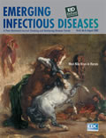
Eugène Delacroix (1798-1863). "Arab Horses Fighting in a Stable." 1860. [PDF - 135 KB - 1 page]
| EID | Potter P. Eugène Delacroix (1798-1863). "Arab Horses Fighting in a Stable." 1860.. Emerg Infect Dis. 2002;8(8):879. https://doi.org/10.3201/eid0808.ac0808 |
|---|---|
| AMA | Potter P. Eugène Delacroix (1798-1863). "Arab Horses Fighting in a Stable." 1860.. Emerging Infectious Diseases. 2002;8(8):879. doi:10.3201/eid0808.ac0808. |
| APA | Potter, P. (2002). Eugène Delacroix (1798-1863). "Arab Horses Fighting in a Stable." 1860.. Emerging Infectious Diseases, 8(8), 879. https://doi.org/10.3201/eid0808.ac0808. |


