Volume 12, Number 6—June 2006
Dispatch
Acanthamoeba Encephalitis in Patient with Systemic Lupus, India
Charudatt G. Shirwadkar*, Rohini Samant*, Milind Sankhe*, Ramesh Deshpande*, Shigeo Yagi†, Frederick L. Schuster†, Rama Sriram‡, and Govinda S. Visvesvara‡
Figure 1
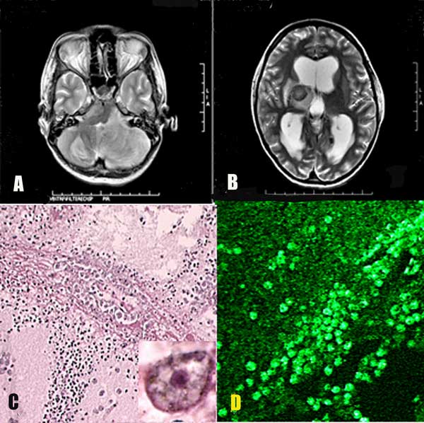
Figure 1. A) Magnetic resonance imaging (MRI) of the patient's brain showing a large lesion in the left cerebellar hemisphere. B) MRI in a different plane taken at the same time. Lesions are evident in the right thalamus and the right half of the pons. C) Blood vessel in brain parenchyma with large numbers of Acanthamoeba in the perivascular space (hematoxylin and eosin stained, magnification ×100). Inset, higher magnification (×1,000) showing nuclear morphology of the ameba. The dark-stained ameba nucleus with a central nucleolus is distinctive. D) Immunofluorescent staining of perivascular brain tissue showing many amebae (magnification ×100).
Page created: January 04, 2012
Page updated: January 04, 2012
Page reviewed: January 04, 2012
The conclusions, findings, and opinions expressed by authors contributing to this journal do not necessarily reflect the official position of the U.S. Department of Health and Human Services, the Public Health Service, the Centers for Disease Control and Prevention, or the authors' affiliated institutions. Use of trade names is for identification only and does not imply endorsement by any of the groups named above.