Volume 15, Number 12—December 2009
Dispatch
Aleutian Mink Disease Virus and Humans
Jørgen R. Jepsen , Francesco d’Amore, Ulrik Baandrup, Michael Roost Clausen, Elisabeth Gottschalck, and Bent Aasted
Figure 1
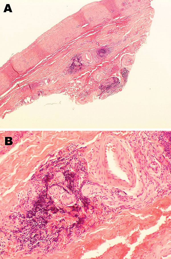
Figure 1. Histopathologic appearance of abdominal aortic biopsy sample from 35-year-old mink farmer in Denmark who had been exposed to Aleutian mink disease parvovirus−infected mink for 10 years (patient 1). A) Perivascular, adventitial lymphoplasmacytoid infiltration. Original magnification ×4. B) Minimal atherosclerotic changes. Original magnification ×20.
Page created: December 09, 2010
Page updated: December 09, 2010
Page reviewed: December 09, 2010
The conclusions, findings, and opinions expressed by authors contributing to this journal do not necessarily reflect the official position of the U.S. Department of Health and Human Services, the Public Health Service, the Centers for Disease Control and Prevention, or the authors' affiliated institutions. Use of trade names is for identification only and does not imply endorsement by any of the groups named above.