Volume 16, Number 3—March 2010
Dispatch
Experimental Infection of Squirrel Monkeys with Nipah Virus
Cite This Article
Citation for Media
Abstract
We infected squirrel monkeys (Saimiri sciureus) with Nipah virus to determine the monkeys’ suitability for use as primate models in preclinical testing of preventive and therapeutic treatments. Infection of squirrel monkeys through intravenous injection was followed by high death rates associated with acute neurologic and respiratory illness and viral RNA and antigen production.
Nipah virus (NiV) is a highly pathogenic zoonotic paramyxovirus that was first identified in Malaysia and Singapore in 1999 (1). Since the initial outbreak, NiV has been associated with human illness in Bangladesh and India (2) and was classified, together with the closely related Hendra virus, in the genus Henipavirus. Reported human death rates varied from 40%–92% (3), and some outbreaks were associated with human-to-human transmission (4). Most human infections led to encephalitis with vasculitis-induced thrombosis in the brain and atypical pneumonia in certain patients (5,6). Because of the lack of efficient treatment or a vaccine for Nipah virus and the high pathogenicity of the virus in humans, the manipulation of NiV requires BioSafety Level 4 (BSL-4) conditions.
Several species of fruit bats of the genus Pteropus are considered natural reservoirs of henipaviruses, although the disease does not develop in them (7). Pigs were responsible for amplifying the NiV infection in Malaysia, but their death rate was only 10%–15%. Laboratory infection of piglets caused development of neurologic signs in some animals, and NiV was detected in different tissues (8). Hamsters in laboratory studies are highly susceptible to NiV, and infection develops in multiple organs, including the brain (9). Cats infected with NiV in the laboratory reproduce the disease observed in naturally infected cats, including a severe respiratory and systemic disease, 6–13 days after infection (10). However, to our knowledge, a primate model necessary for preclinical testing of preventive and therapeutic approaches has not been described. We therefore assessed the squirrel monkey (Saimiri sciureus) as an experimental model of NiV infection.
We selected these New World monkeys because of their availability, reliability as a primate model with which to study infectious diseases (11), and suitability as experimental animals in BSL-4 conditions. Thirteen 4-year-old male monkeys (0.8–1.0 kg) were imported from a breeding colony in French Guiana and housed in the BSL-4 animal care facility in Lyon. Experimental methods were approved by the Région Rhône Alpes ethics committee.
Twelve monkeys were infected with NiV isolate UMMC1 (1), GenBank accession no. AY029767, either intravenously or intranasally; for both modes of infection either 103 or 107 PFU was used. Animals were observed daily for 2 months for signs of disease onset; tissues were taken during the infection and at necropsy or at the end of experiment (Table 1). Blood samples were collected at different time points, serum samples were used for antibody analysis, and peripheral blood cells (PBMC) were used for RNA isolation. Different organ samples were taken and frozen at –80°C for RNA isolation or fixed in 4% formalin for histopathologic studies.
RNA was extracted from different organs and analyzed by 1-step RT-PCR by using high fidelity PCR enzyme blend (Roche Applied Science, Mannheim, Germany) for NiV nucleoprotein expression as described (12). Detection of NiV-specific antibodies in the serum was performed simultaneously for all samples by ELISA and virus neutralization assays as described (13). Immunohistochemical analysis was conducted on formalin-fixed, paraffin-embedded tissues as described (6).
Onset of clinical illness was observed between 7 and 19 days postinfection (dpi), with development in the animals of anorexia, weight loss, and depression (characterized by slumped, collapsed body posture and lack of responsiveness to the environmental triggers). These clinical signs progressed for several hours and were associated with hyperthermia and an acute respiratory syndrome characterized by dyspnea and hyperventilation. During the course of the disease, the animals became more obtunded and had uncoordinated motor movements, ending, in some instances, with a loss of consciousness and coma (Table 1). Although clinical signs were seen in monkeys infected intranasally and intravenously, the disease lasted longer in intranasally infected animals (7 days) than in intravenously infected monkeys (2–3 days). With the latter, death was observed in 3 of 4 animals in which the disease was allowed to proceed. Clinical signs of illness for intranasally infected monkeys were milder and seen only in 2 of 4 animals before recovery after 3–7 days of illness. Clinical signs observed in monkeys appear to be similar to those reported for human infection, including involvement of neurologic and respiratory systems. In addition, the incubation period for the acute human infection in Malaysia was estimated to be from a few days to 2 weeks, total duration of illness ranged 2–34 days, and the rate of subclinical infection was ≈25% (6,14). It is possible that the inclusion of more animals in the study would have given higher heterogeneity in the course of disease, as seen in humans. Intravenous infection was much more efficient than the intranasal route in monkeys, probably because of a better delivery of the virus to different tissues.
NiV-specific RNA was detected in various organs only in intravenously infected animals (Appendix Table), demonstrating a differential virus spread, depending on time after infection and virus dose. Early detection after infection (3 dpi) was possible only in animal D, which was infected with a high dose of NiV. Animal F, which recovered from the disease, although positive for NiV (by RT-PCR) in the PBMC sample 2 dpi, was negative after necropsy on day 52, when virus was probably eliminated from the monkey. Detection of viral RNA in different tissues (liver, brain, spleen, kidney, lung, lymph nodes) early after infection suggests a rapid propagation of NiV and tropism for various tissues. Viral RNA was found in PBMC taken at different time points after infection, suggesting the role of these cells in viral propagation in the monkey. In contrast to what has been observed in hamsters (9), viral RNA was not detected in any urine samples from analyzed animals, thus excluding urine as a possible mode of virus dissemination in this species.
Figure
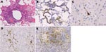
Figure. Pathologic signs associated with Nipah virus infection in squirrel monkeys. A) Focal inflammation in the lung (monkey B). Hematoxylin and eosin stains; original magnification ×10. B) Viral antigens (brown staining) were...
Monkeys showed mild histologic lesions, including the inflammation most obvious in the lung parenchyma (Figure, panel A). In contrast to human infection, vasculitis and brain abnormalities were much less evident. However, immunohistochemistry showed viral antigens immunolocalized to the brain, lung, spleen, and kidney extravascular parenchyma, thus confirming viral infection in these organs (Figure, panels B–E).
NiV-specific immunoglobulin (Ig) M responses were observed starting from 8 dpi for all monkeys except in groups H and I (Table 2). This finding suggests that 103 PFU of NiV delivered intranasally was probably insufficient to induce infection in monkeys. Although NiV-specific antibodies were detected by ELISA in animals dying from the infection, sufficient titers of neutralizing antibodies did not develop in these monkeys and they were therefore not protected. These findings suggest the protective role of high neutralization titers in NiV infection. Our results agree with other studies of NiV infection that reported most human patients with fatal NiV infection had IgG and IgM in their serum and cerebrospinal fluid (6,15); neutralization titers were not analyzed in those studies.
Our results suggest some similarities of NiV pathogenesis in humans and squirrel monkeys, including development of clinical signs, progression of infection, and humoral immune response. We conclude that the squirrel monkey can be used as an animal model for experimental studies of NiV infection, and these results pave the way for further study.
Dr Marianneau is a senior scientist and project leader for flaviviruses and BSL-4 viruses at Institut Pasteur. His research is focused on the physiopathology of hemorrhagic fever viruses and other BSL-4 agents.
Acknowledgments
We thank BioSafety team members from BSL-4 Laboratory “Jean Mérieux” for their assistance.
The work was supported by Institut Pasteur, Institut national de santé et de la recherché médicale, ANR 2005 program “Microbologie, infections et immunités” (ANR-05-MIIM-017-02), and the government of Malaysia (IRPA 06-02-03-0000-PR0060/04).
References
- Chan YP, Chua KB, Koh CL, Lim ME, Lam SK. Complete nucleotide sequences of Nipah virus isolates from Malaysia. J Gen Virol. 2001;82:2151–5.PubMedGoogle Scholar
- Harcourt BH, Lowe L, Tamin A, Liu X, Bankamp B, Bowden N, Genetic characterization of Nipah virus, Bangladesh, 2004. Emerg Infect Dis. 2005;10:1594–7.
- Luby SP, Rahman M, Hossain MJ, Blum LS, Husain MM, Gurley E, Foodborne transmission of Nipah virus, Bangladesh. Emerg Infect Dis. 2006;12:1888–94.PubMedGoogle Scholar
- Gurley ES, Montgomery J, Hossain MJ, Bell M, Azad AK, Islam MR, Person-to-person transmission of Nipah virus in a Bangladeshi community. Emerg Infect Dis. 2007;13:1031–7.PubMedGoogle Scholar
- Chua KB, Goh KJ, Wong KT, Kamarulzaman A, Tan PS, Ksiazek TG, Fatal encephalitis due to Nipah virus among pig-farmers in Malaysia. Lancet. 1999;354:1257–9. DOIPubMedGoogle Scholar
- Wong KT, Shieh WJ, Kumar S, Norain K, Abdullah W, Guarner J, Nipah virus infection: pathology and pathogenesis of an emerging paramyxoviral zoonosis. Am J Pathol. 2002;161:2153–67.PubMedGoogle Scholar
- Chua KB, Koh CL, Hooi PS, Wee KF, Khong JH, Chua BH, Isolation of Nipah virus from Malaysian island flying-foxes. Microbes Infect. 2002;4:145–51. DOIPubMedGoogle Scholar
- Weingartl H, Czub S, Copps J, Berhane Y, Middleton D, Marszal P, Invasion of the central nervous system in a porcine host by Nipah virus. J Virol. 2005;79:7528–34. DOIPubMedGoogle Scholar
- Wong KT, Grosjean I, Brisson C, Blanquier B, Fevre-Montange M, Bernard A, A golden hamster model for human acute Nipah virus infection. Am J Pathol. 2003;163:2127–37.PubMedGoogle Scholar
- Mungall BA, Middleton D, Crameri G, Bingham J, Halpin K, Russell G, Feline model of acute Nipah virus infection and protection with a soluble glycoprotein-based subunit vaccine. J Virol. 2006;80:12293–302. DOIPubMedGoogle Scholar
- Contamin H, Behr C, Mercereau-Puijalon O, Michel J. Plasmodium falciparum in the squirrel monkey (Saimiri sciureus): infection of non-splenectomised animals as a model for exploring clinical manifestations of malaria. Microbes Infect. 2000;2:945–54. DOIPubMedGoogle Scholar
- Guillaume V, Contamin H, Loth P, Grosjean I, Courbot MC, Deubel V, Antibody prophylaxis and therapy against Nipah virus infection in hamsters. J Virol. 2006;80:1972–8. DOIPubMedGoogle Scholar
- Guillaume V, Wong KT, Looi RY, Georges-Courbot MC, Barrot L, Buckland R, Acute Hendra virus infection: analysis of the pathogenesis and passive antibody protection in the hamster model. Virology. 2009;387:459–65. DOIPubMedGoogle Scholar
- Chadha MS, Comer JA, Lowe L, Rota PA, Rollin PE, Bellini WJ, Nipah virus-associated encephalitis outbreak, Siliguri, India. Emerg Infect Dis. 2006;12:235–40.PubMedGoogle Scholar
Figure
Tables
Cite This ArticleTable of Contents – Volume 16, Number 3—March 2010
| EID Search Options |
|---|
|
|
|
|
|
|
Please use the form below to submit correspondence to the authors or contact them at the following address:
Branka Horvat, Institut national de santé et de la recherché médicale, U758, 21 ave T. Garnier, Lyon 69007, France
Top