Volume 20, Number 9—September 2014
Letter
Cerebellitis Associated with Influenza A(H1N1)pdm09, United States, 2013
Maroun M. Sfeir and Catherine E. Najem
Figure
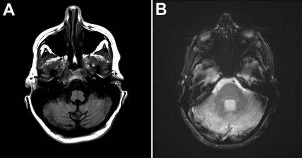
Figure. A) Magnetic resonance images of the brain of a woman with cerebellitis associated with influenza A(H1N1)pdm09, United States, 2013. T1-weighted axial MRI brain sequence showing hypo-intensity of bilateral cerebellar hemispheres. B) T2-weighted axial MRI brain sequence showing hyperintensity of bilateral cerebellar hemispheres.
Page created: August 18, 2014
Page updated: August 18, 2014
Page reviewed: August 18, 2014
The conclusions, findings, and opinions expressed by authors contributing to this journal do not necessarily reflect the official position of the U.S. Department of Health and Human Services, the Public Health Service, the Centers for Disease Control and Prevention, or the authors' affiliated institutions. Use of trade names is for identification only and does not imply endorsement by any of the groups named above.