Volume 21, Number 9—September 2015
CME ACTIVITY - Synopsis
Mycobacterium abscessus Complex Infections in Humans
Meng-Rui Lee, Wang-Huei Sheng, Chien-Ching Hung, Chong-Jen Yu, Li-Na Lee, and Po-Ren Hsueh
Figure 5
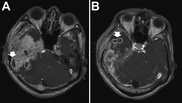
Figure 5. Brain computed tomography scan images for a patient with central nervous system infection caused by Mycobacterium abscessus subsp. bolletii. Arrows indicate abnormal nodular pachymeningeal thickening and leptomeningeal and intraparenchymal extension with multiple rim-enhancing lesions in the right cerebellum (A) and right temporal lobe (B), indicating cerebral abscesses.
Page created: August 14, 2015
Page updated: August 14, 2015
Page reviewed: August 14, 2015
The conclusions, findings, and opinions expressed by authors contributing to this journal do not necessarily reflect the official position of the U.S. Department of Health and Human Services, the Public Health Service, the Centers for Disease Control and Prevention, or the authors' affiliated institutions. Use of trade names is for identification only and does not imply endorsement by any of the groups named above.