Volume 22, Number 4—April 2016
Dispatch
Cross-Neutralization between Human and African Bat Mumps Viruses
Figure 1

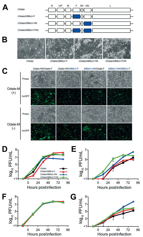
Figure 1. Construction of rMuVs expressing the ABMuV envelope proteins for study of their functions. A) Genome structures of the rMuVs. The 7 boxes indicate the N, V/P, M, F, SH, HN, and L genes of MuV. The blue boxes indicate the genes derived from ABMuV. B) Cytopathic effect of rMuV infection of the Vero cells followed by incubation for 48 hr. C) BHK cells were transfected with expression plasmids of the HN and F proteins (pCAGGS-Odate-HN or -ABMuV-HN and pCAGGS-Odate-F or -ABMuV-F). They were also cotransfected with expression plasmids of the M protein (pCAGGS-Odate-M) and AcGFP (pAcGFP-C1, Clontech). At 48 h posttransfection, the cells were observed under a phase-contrast and a fluorescence microscope. D–G) Growth kinetics of the rMuVs in Vero (African green monkey) (D), A549 (human) (E), THP-1 (human) (F) and FBKT1 (Ryukyu fruit bat) (G) cells. Each cell line was infected with the rMuVs at a multiplicity of infection of 0.01. At the indicated times postinfection, the culture supernatants were collected, and the infectious titers were determined by plaque assay. ABMuV, African bat mumps virus; F, fusion; HN, hemagglutinin-neuraminidase; L, large; M, matrix; N, nucleocapsid; rMuV, recombinant mumps virus; SH, small hydrophobic; P, phosphoprotein; V, historically considered the fifth viral protein. Error bars indicate SD.