Volume 9, Number 3—March 2003
Synopsis
Electron Microscopy for Rapid Diagnosis of Emerging Infectious Agents1
Figure 6
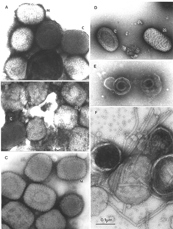
Figure 6. A–EComparison of clinically relevant viral agents associated with skin lesionsA–C show poxviruses indistinguishable in appearance from variola virus, the agent of smallpoxThe slightly rounded, brick-shaped virions measure about 270 by 350 nmTwo types of particles may be seenM, or Mulberry forms show a 10- to 20-nm diameter short-tubular or beaded surface (M)Capsular, or C forms, partly penetrated by the stain, are recognized by a 30-nm membrane (C): AMolluscum contagiosum (molluscipoxvirus) virions from skin lesions observed in an adult; BVaccinia virus vaccine strain WR (orthopoxvirus) from cell culture; CEctromelia virus (orthopoxvirus) from culture materialDParapox viruses measure up to190 by 300 nm and are more distinctly ovoidTubules, 10 to 20 nm wide and approximately 1,000-nm long, spiral around the virion, giving a distinctive crosshatched appearanceEHerpesvirus particles from a skin lesion of a primary varicella zoster infection observed in an adultDirect electron microscopic shows two virionsThe envelopes are broken, liberating the 100-nm nucleocapsidFCell culture supernatant from a patient with an infantile respiratory tract infectionThe enveloped virions are studded with tiny surface spikesThe 18-nm helical nucleocapsids have been released from disintegrating virionsThe nucleocapsids and envelope details are typical of paramyxovirusesA–B, phosphotungstic acid, C–F, uranyl acetateAll prints at the same magnification, bar = 100 nm.