Perspective
Anthrax of the Gastrointestinal Tract [PDF - 237 KB - 3 pages]
When swallowed, anthrax spores may cause lesions from the oral cavity to the cecum. Gastrointestinal anthrax is greatly underreported in rural disease-endemic areas of the world. The apparent paucity of this form of anthrax reflects the lack of facilities able to make the diagnosis in these areas. The spectrum of disease, ranging from subclinical infection to death, has not been fully recognized. In some community-based studies, cases of gastrointestinal anthrax outnumbered those of cutaneous anthrax. The oropharyngeal variant, in particular, is unfamiliar to most physicians. The clinical features of oropharyngeal anthrax include fever and toxemia, inflammatory lesion(s) in the oral cavity or oropharynx, enlargement of cervical lymph nodes associated with edema of the soft tissue of the cervical area, and a high case-fatality rate. Awareness of gastrointestinal anthrax in a differential diagnosis remains important in anthrax-endemic areas but now also in settings of possible bioterrorism.
| EID | Sirisanthana T, Brown AE. Anthrax of the Gastrointestinal Tract. Emerg Infect Dis. 2002;8(7):649-651. https://doi.org/10.3201/eid0807.020062 |
|---|---|
| AMA | Sirisanthana T, Brown AE. Anthrax of the Gastrointestinal Tract. Emerging Infectious Diseases. 2002;8(7):649-651. doi:10.3201/eid0807.020062. |
| APA | Sirisanthana, T., & Brown, A. E. (2002). Anthrax of the Gastrointestinal Tract. Emerging Infectious Diseases, 8(7), 649-651. https://doi.org/10.3201/eid0807.020062. |
Research
Emergence of Usutu virus, an African Mosquito-Borne Flavivirus of the Japanese Encephalitis Virus Group, Central Europe [PDF - 942 KB - 5 pages]
During late summer 2001 in Austria, a series of deaths in several species of birds occurred, similar to the beginning of the West Nile virus (WNV) epidemic in the United States. We necropsied the dead birds and examined them by various methods; pathologic and immunohistologic investigations suggested a WNV infection. Subsequently, the virus was isolated, identified, partially sequenced, and subjected to phylogenetic analysis. The isolates exhibited 97% identity to Usutu virus (USUV), a mosquito-borne Flavivirus of the Japanese encephalitis virus group; USUV has never previously been observed outside Africa nor associated with fatal disease in animals or humans. If established in central Europe, this virus may have considerable effects on avian populations; whether USUV has the potential to cause severe human disease is unknown.
| EID | Weissenböck H, Kolodziejek J, Url A, Lussy H, Rebel-Bauder B, Nowotny N. Emergence of Usutu virus, an African Mosquito-Borne Flavivirus of the Japanese Encephalitis Virus Group, Central Europe. Emerg Infect Dis. 2002;8(7):652-656. https://doi.org/10.3201/eid0807.020094 |
|---|---|
| AMA | Weissenböck H, Kolodziejek J, Url A, et al. Emergence of Usutu virus, an African Mosquito-Borne Flavivirus of the Japanese Encephalitis Virus Group, Central Europe. Emerging Infectious Diseases. 2002;8(7):652-656. doi:10.3201/eid0807.020094. |
| APA | Weissenböck, H., Kolodziejek, J., Url, A., Lussy, H., Rebel-Bauder, B., & Nowotny, N. (2002). Emergence of Usutu virus, an African Mosquito-Borne Flavivirus of the Japanese Encephalitis Virus Group, Central Europe. Emerging Infectious Diseases, 8(7), 652-656. https://doi.org/10.3201/eid0807.020094. |
First Human Isolate of Hantavirus (Andes virus) in the Americas [PDF - 1018 KB - 5 pages]
We isolated Andes virus (formal name: Andes virus [ANDV], a species in the genus Hantavirus), from serum of an asymptomatic 10-year-old Chilean boy who died 6 days later of hantavirus pulmonary syndrome (HPS). The serum was obtained 12 days after his grandmother died from HPS and 2 days before he became febrile. No hantavirus immunoglobulin (Ig) G or IgM antibodies were detected in the serum sample. After three blind passages, ANDV antigens were detected in Vero E6 cells by immunofluorescence assay and enzyme-linked immunosorbent assay, and ANDV RNA was detected by reverse transcription-polymerase chain reaction. A fragment of the virus genome showed 96.2% nucleotide identity with that of prototype ANDV. To our knowledge, this is the first isolation of any agent of hemorrhagic fever with renal syndrome from a human and the first such isolation of hantavirus before symptoms of that syndrome or HPS began.
| EID | Galeno H, Mora J, Villagra E, Fernandez J, Hernandez J, Mertz GJ, et al. First Human Isolate of Hantavirus (Andes virus) in the Americas. Emerg Infect Dis. 2002;8(7):657-661. https://doi.org/10.3201/eid0807.010277 |
|---|---|
| AMA | Galeno H, Mora J, Villagra E, et al. First Human Isolate of Hantavirus (Andes virus) in the Americas. Emerging Infectious Diseases. 2002;8(7):657-661. doi:10.3201/eid0807.010277. |
| APA | Galeno, H., Mora, J., Villagra, E., Fernandez, J., Hernandez, J., Mertz, G. J....Ramirez, E. (2002). First Human Isolate of Hantavirus (Andes virus) in the Americas. Emerging Infectious Diseases, 8(7), 657-661. https://doi.org/10.3201/eid0807.010277. |
Ecologic Niche Modeling and Potential Reservoirs for Chagas Disease, Mexico. [PDF - 1.30 MB - 6 pages]
Ecologic niche modeling may improve our understanding of epidemiologically relevant vector and parasite-reservoir distributions. We used this tool to identify host relationships of Triatoma species implicated in transmission of Chagas disease. Associations have been documented between the protracta complex (Triatoma: Triatominae: Reduviidae) with packrat species (Neotoma spp.), providing an excellent case study for the broader challenge of developing hypotheses of association. Species pairs that were identified coincided exactly with those in previous studies, suggesting that local interactions between Triatoma and Neotoma species and subspecies have implications at a geographic level. Nothing is known about sylvatic associates of T. barberi, which are considered the primary Chagas vector in Mexico; its geographic distribution coincided closely with that of N. mexicana, suggesting interaction. The presence of the species was confirmed in two regions where it had been predicted but not previously collected. This approach may help in identifying Chagas disease risk areas, planning vector-control strategies, and exploring parasite-reservoir associations for other emerging diseases.
| EID | Peterson AT, Sánchez-Cordero V, Ben Beard C, Ramsey JM. Ecologic Niche Modeling and Potential Reservoirs for Chagas Disease, Mexico.. Emerg Infect Dis. 2002;8(7):662-667. https://doi.org/10.3201/eid0807.010454 |
|---|---|
| AMA | Peterson AT, Sánchez-Cordero V, Ben Beard C, et al. Ecologic Niche Modeling and Potential Reservoirs for Chagas Disease, Mexico.. Emerging Infectious Diseases. 2002;8(7):662-667. doi:10.3201/eid0807.010454. |
| APA | Peterson, A. T., Sánchez-Cordero, V., Ben Beard, C., & Ramsey, J. M. (2002). Ecologic Niche Modeling and Potential Reservoirs for Chagas Disease, Mexico.. Emerging Infectious Diseases, 8(7), 662-667. https://doi.org/10.3201/eid0807.010454. |
Ehrlichia ewingii Infection in White-Tailed Deer (Odocoileus virginianus)
Two closely related zoonotic ehrlichiae, Ehrlichia chaffeensis and E. ewingii, are transmitted by Amblyomma americanum, the lone star tick. Because white-tailed deer (Odocoileus virginianus) are critical hosts for all mobile stages of A. americanum and are important vertebrate reservoirs of E. chaffeensis, we investigated whether deer may be infected with E. ewingii, a cause of granulocytotropic ehrlichiosis in humans and dogs. To test for E. ewingii infection, we used polymerase chain reaction and inoculation of fawns with whole blood from wild deer. Of 110 deer tested from 20 locations in 8 U.S. states, 6 (5.5%) were positive for E. ewingii. In addition, natural E. ewingii infection was confirmed through infection of captive fawns. These findings expand the geographic distribution of E. ewingii, along with risk for human infection, to include areas of Kentucky, Georgia, and South Carolina. These data suggest that white-tailed deer may be an important reservoir for E. ewingii.
| EID | Yabsley MJ, Varela AS, Tate CM, Dugan VG, Stallknecht DE, Little SE, et al. Ehrlichia ewingii Infection in White-Tailed Deer (Odocoileus virginianus). Emerg Infect Dis. 2002;8(7):668-671. https://doi.org/10.3201/eid0807.020018 |
|---|---|
| AMA | Yabsley MJ, Varela AS, Tate CM, et al. Ehrlichia ewingii Infection in White-Tailed Deer (Odocoileus virginianus). Emerging Infectious Diseases. 2002;8(7):668-671. doi:10.3201/eid0807.020018. |
| APA | Yabsley, M. J., Varela, A. S., Tate, C. M., Dugan, V. G., Stallknecht, D. E., Little, S. E....Davidson, W. R. (2002). Ehrlichia ewingii Infection in White-Tailed Deer (Odocoileus virginianus). Emerging Infectious Diseases, 8(7), 668-671. https://doi.org/10.3201/eid0807.020018. |
Time-Space Clustering of Human Brucellosis, California, 1973–1992 [PDF - 431 KB - 7 pages]
Infection with Brucella spp. continues to pose a human health risk in California despite great strides in eradicating the disease from domestic animals. Clustering of human cases in time and space has important public health implications for understanding risk factors and sources of infection. Temporal-spatial clustering of human brucellosis in California for the 20-year period 1973–1992 was evaluated by the Ederer-Myers-Mantel, Moran’s I, and population-adjusted Moran’s I procedures. Cases were clustered in concentrated agricultural regions in the first 5-year interval (1973–1977). Time-space clustering of human brucellosis cases in California late in the 20-year study period may reflect the distribution of Hispanic populations. Public health programs in California should focus on educating Hispanic populations about the risk of consuming dairy products, such as soft cheeses, made from unpasteurized milk.
| EID | Fosgate GT, Carpenter TE, Chomel BB, Case JT, DeBess EE, Reilly KF. Time-Space Clustering of Human Brucellosis, California, 1973–1992 . Emerg Infect Dis. 2002;8(7):672-678. https://doi.org/10.3201/eid0807.010351 |
|---|---|
| AMA | Fosgate GT, Carpenter TE, Chomel BB, et al. Time-Space Clustering of Human Brucellosis, California, 1973–1992 . Emerging Infectious Diseases. 2002;8(7):672-678. doi:10.3201/eid0807.010351. |
| APA | Fosgate, G. T., Carpenter, T. E., Chomel, B. B., Case, J. T., DeBess, E. E., & Reilly, K. F. (2002). Time-Space Clustering of Human Brucellosis, California, 1973–1992 . Emerging Infectious Diseases, 8(7), 672-678. https://doi.org/10.3201/eid0807.010351. |
Persistent High Incidence of Tuberculosis in Immigrants in a Low-Incidence Country [PDF - 216 KB - 6 pages]
Immigration from areas of high incidence is thought to have fueled the resurgence of tuberculosis (TB) in areas of low incidence. To reduce the risk of disease in low-incidence areas, the main countermeasure has been the screening of immigrants on arrival. This measure is based on the assumption of a prompt decline in the incidence of TB in immigrants during their first few years of residence in a country with low overall incidence. We have documented that this assumption is not true for 619 Somali immigrants reported in Denmark as having TB. The annual incidence of TB declined only gradually during the first 7 years of residence, from an initial 2,000 per 100,000 to 700 per 100,000. The decline was described by an exponential function with a half-time of 5.7 (95% confidence interval 4.0 to 9.7) years. This finding seriously challenges the adequacy of the customary practice of screening solely on arrival.
| EID | Lillebaek T, Andersen ÅB, Dirksen A, Smith E, Skovgaard LT, Kok-Jensen A. Persistent High Incidence of Tuberculosis in Immigrants in a Low-Incidence Country. Emerg Infect Dis. 2002;8(7):679-684. https://doi.org/10.3201/eid0807.010482 |
|---|---|
| AMA | Lillebaek T, Andersen ÅB, Dirksen A, et al. Persistent High Incidence of Tuberculosis in Immigrants in a Low-Incidence Country. Emerging Infectious Diseases. 2002;8(7):679-684. doi:10.3201/eid0807.010482. |
| APA | Lillebaek, T., Andersen, Å. B., Dirksen, A., Smith, E., Skovgaard, L. T., & Kok-Jensen, A. (2002). Persistent High Incidence of Tuberculosis in Immigrants in a Low-Incidence Country. Emerging Infectious Diseases, 8(7), 679-684. https://doi.org/10.3201/eid0807.010482. |
Automatic Electronic Laboratory-Based Reporting of Notifiable Infectious Diseases [PDF - 293 KB - 7 pages]
Electronic laboratory-based reporting, developed by the University of Pittsburgh Medical Center (UPMC) Health System, was evaluated to determine if it could be integrated into the conventional paper-based reporting system. We reviewed reports of 10 infectious diseases from 8 UPMC hospitals that reported to the Allegheny County Health Department in southwestern Pennsylvania during January 1–November 26, 2000. Electronic reports were received a median of 4 days earlier than conventional reports. The completeness of reporting was 74% (95% confidence interval [CI] 66% to 81%) for the electronic laboratory-based reporting and 65% (95% CI 57% to 73%) for the conventional paper-based reporting system (p>0.05). Most reports (88%) missed by electronic laboratory-based reporting were caused by using free text. Automatic reporting was more rapid and as complete as conventional reporting. Using standardized coding and minimizing free text usage will increase the completeness of electronic laboratory-based reporting.
| EID | Panackal AA, Tsui F, McMahon J, Wagner MM, Dixon BW, Zubieta J, et al. Automatic Electronic Laboratory-Based Reporting of Notifiable Infectious Diseases. Emerg Infect Dis. 2002;8(7):685-691. https://doi.org/10.3201/eid0807.010493 |
|---|---|
| AMA | Panackal AA, Tsui F, McMahon J, et al. Automatic Electronic Laboratory-Based Reporting of Notifiable Infectious Diseases. Emerging Infectious Diseases. 2002;8(7):685-691. doi:10.3201/eid0807.010493. |
| APA | Panackal, A. A., Tsui, F., McMahon, J., Wagner, M. M., Dixon, B. W., Zubieta, J....Harrison, L. H. (2002). Automatic Electronic Laboratory-Based Reporting of Notifiable Infectious Diseases. Emerging Infectious Diseases, 8(7), 685-691. https://doi.org/10.3201/eid0807.010493. |
Infection by Ralstonia Species in Cystic Fibrosis Patients: Identification of R. pickettii and R. mannitolilytica by Polymerase Chain Reaction [PDF - 367 KB - 5 pages]
The frequency of respiratory tract infections caused by Ralstonia species in persons with cystic fibrosis (CF) and the role of these species in CF pulmonary disease are not well documented. In part, this lack of documentation may be attributed to the difficulty in accurately identifying Ralstonia species; R. mannitolilytica and R. pickettii in particular may be misidentified as other closely related species, particularly those of the Burkholderia cepacia complex. We used polyphasic analyses to identify 42 Ralstonia isolates from sputum cultures from 38 CF patients. Several isolates that could not be identified to the species level may belong to novel Ralstonia species. We demonstrated chronic colonization by using genotyping of serial isolates recovered from the same patient. To facilitate identification of R. mannitolilytica and R. pickettii, we developed 16S ribosomal DNA-based polymerase chain reaction assays that allow sensitive and specific identification of these species.
| EID | Coenye T, Vandamme P, LiPuma JJ. Infection by Ralstonia Species in Cystic Fibrosis Patients: Identification of R. pickettii and R. mannitolilytica by Polymerase Chain Reaction. Emerg Infect Dis. 2002;8(7):692-696. https://doi.org/10.3201/eid0807.010472 |
|---|---|
| AMA | Coenye T, Vandamme P, LiPuma JJ. Infection by Ralstonia Species in Cystic Fibrosis Patients: Identification of R. pickettii and R. mannitolilytica by Polymerase Chain Reaction. Emerging Infectious Diseases. 2002;8(7):692-696. doi:10.3201/eid0807.010472. |
| APA | Coenye, T., Vandamme, P., & LiPuma, J. J. (2002). Infection by Ralstonia Species in Cystic Fibrosis Patients: Identification of R. pickettii and R. mannitolilytica by Polymerase Chain Reaction. Emerging Infectious Diseases, 8(7), 692-696. https://doi.org/10.3201/eid0807.010472. |
Temporal Changes in Prevalence of Antimicrobial Resistance in 23 U.S. Hospitals [PDF - 215 KB - 5 pages]
Antimicrobial resistance is increasing in nearly all health-care–associated pathogens. We examined changes in resistance prevalence during 1996–1999 in 23 hospitals by using two statistical methods. When the traditional chi-square test of pooled mean resistance prevalence was used, most organisms appear to have increased in prevalence. However, when a more conservative test that accounts for changes within individual hospitals was used, significant increases in prevalence of resistance were consistently observed only for oxacillin-resistant Staphylococcus aureus, ciprofloxacin-resistant Pseudomonas aeruginosa, and ciprofloxacin- or ofloxacin-resistant Escherichia coli. These increases were significant only in isolates from patients outside intensive-care units (ICU). The increases seen are of concern; differences in factors present outside ICUs, such as excessive quinolone use or inadequate infection-control practices, may explain the observed trends.
| EID | Hospitals P, Fridkin SK, Hill HA, Volkova NV, Edwards JR, Lawton RM, et al. Temporal Changes in Prevalence of Antimicrobial Resistance in 23 U.S. Hospitals. Emerg Infect Dis. 2002;8(7):697-701. https://doi.org/10.3201/eid0807.010427 |
|---|---|
| AMA | Hospitals P, Fridkin SK, Hill HA, et al. Temporal Changes in Prevalence of Antimicrobial Resistance in 23 U.S. Hospitals. Emerging Infectious Diseases. 2002;8(7):697-701. doi:10.3201/eid0807.010427. |
| APA | Hospitals, P., Fridkin, S. K., Hill, H. A., Volkova, N. V., Edwards, J. R., Lawton, R. M....McGowan, J. E. (2002). Temporal Changes in Prevalence of Antimicrobial Resistance in 23 U.S. Hospitals. Emerging Infectious Diseases, 8(7), 697-701. https://doi.org/10.3201/eid0807.010427. |
Monitoring Antimicrobial Use and Resistance: Comparison with a National Benchmark on Reducing Vancomycin Use and Vancomycin-Resistant Enterococci [PDF - 249 KB - 6 pages]
To determine if local monitoring data on vancomycin use directed quality improvement and decreased vancomycin use or vancomycin-resistant enterococci (VRE), we analyzed data from 50 intensive-care units (ICUs) at 20 U.S. hospitals reporting data on antimicrobial-resistant organisms and antimicrobial agent use. We compared local data with national benchmark data (aggregated from all study hospitals). After data were adjusted for changes in prevalence of methicillin-resistant Staphylococcus aureus, changes in specific prescriber practice at ICUs were associated with significant decreases in vancomycin use (mean decrease -48 defined daily doses per 1,000 patient days, p<0.001). These ICUs also reported significant decreases in VRE prevalence compared with those not using unit-specific changes in practice (mean decrease of 7.5% compared with mean increase of 5.7%, p<0.001). In this study, practice changes focused towards specific ICUs were associated with decreases in ICU vancomycin use and VRE prevalence.
| EID | Fridkin SK, Lawton R, Edwards JR, Tenover FC, McGowan JE, Gaynes RP. Monitoring Antimicrobial Use and Resistance: Comparison with a National Benchmark on Reducing Vancomycin Use and Vancomycin-Resistant Enterococci. Emerg Infect Dis. 2002;8(7):702-707. https://doi.org/10.3201/eid0807.010465 |
|---|---|
| AMA | Fridkin SK, Lawton R, Edwards JR, et al. Monitoring Antimicrobial Use and Resistance: Comparison with a National Benchmark on Reducing Vancomycin Use and Vancomycin-Resistant Enterococci. Emerging Infectious Diseases. 2002;8(7):702-707. doi:10.3201/eid0807.010465. |
| APA | Fridkin, S. K., Lawton, R., Edwards, J. R., Tenover, F. C., McGowan, J. E., & Gaynes, R. P. (2002). Monitoring Antimicrobial Use and Resistance: Comparison with a National Benchmark on Reducing Vancomycin Use and Vancomycin-Resistant Enterococci. Emerging Infectious Diseases, 8(7), 702-707. https://doi.org/10.3201/eid0807.010465. |
Prevalence, Distribution, and Host Range of Peste des petits ruminants virus, Turkey [PDF - 296 KB - 4 pages]
Peste des petits ruminants virus (PPRV, genus Morbillivirus), which causes a severe disease in sheep and goats, has only recently been officially declared to be present in Turkey. We carried out a study to determine the prevalence, distribution, and host range of PPRV in Turkey. A total of 1,607 animals, reared in 18 different locations, were monitored for the presence of antibodies to PPRV and the related virus of large ruminants, Rinderpest virus (RPV). Only two farms had animals that were free of antibody responses to either disease. Prevalence for PPRV infection varied (range 0.87%–82.6%) and was higher in sheep (29.2%) than in goats (20%). The overall antibody responses to PPRV and RPV were 22.4% and 6.28%, respectively. Two PPRVs of lineage 4, which comprises many other PPRVs whose origins are in the Middle East, the Arabian Peninsula, and southern Asia, were isolated from Turkish sheep.
| EID | Özkul A, Akca Y, Alkan F, Barrett T, Karaoglu T, Dagalp SB, et al. Prevalence, Distribution, and Host Range of Peste des petits ruminants virus, Turkey. Emerg Infect Dis. 2002;8(7):709-712. https://doi.org/10.3201/eid0807.010471 |
|---|---|
| AMA | Özkul A, Akca Y, Alkan F, et al. Prevalence, Distribution, and Host Range of Peste des petits ruminants virus, Turkey. Emerging Infectious Diseases. 2002;8(7):709-712. doi:10.3201/eid0807.010471. |
| APA | Özkul, A., Akca, Y., Alkan, F., Barrett, T., Karaoglu, T., Dagalp, S. B....Burgu, I. (2002). Prevalence, Distribution, and Host Range of Peste des petits ruminants virus, Turkey. Emerging Infectious Diseases, 8(7), 709-712. https://doi.org/10.3201/eid0807.010471. |
Antibiotic Resistance of Gram-Negative Bacteria in Rivers, United States [PDF - 207 KB - 4 pages]
Bacteria with intrinsic resistance to antibiotics are found in nature. Such organisms may acquire additional resistance genes from bacteria introduced into soil or water, and the resident bacteria may be the reservoir or source of widespread resistant organisms found in many environments. We isolated antibiotic-resistant bacteria in freshwater samples from 16 U.S. rivers at 22 sites and measured the prevalence of organisms resistant to β-lactam and non β-lactam antibiotics. Over 40% of the bacteria resistant to more than one antibiotic had at least one plasmid. Ampicillin resistance genes, as well as other resistance traits, were identified in 70% of the plasmids. The most common resistant organisms belonged to the following genera: Acinetobacter, Alcaligenes, Citrobacter, Enterobacter, Pseudomonas, and Serratia.
| EID | Ash RJ, Mauck B, Morgan M. Antibiotic Resistance of Gram-Negative Bacteria in Rivers, United States. Emerg Infect Dis. 2002;8(7):713-716. https://doi.org/10.3201/eid0807.010264 |
|---|---|
| AMA | Ash RJ, Mauck B, Morgan M. Antibiotic Resistance of Gram-Negative Bacteria in Rivers, United States. Emerging Infectious Diseases. 2002;8(7):713-716. doi:10.3201/eid0807.010264. |
| APA | Ash, R. J., Mauck, B., & Morgan, M. (2002). Antibiotic Resistance of Gram-Negative Bacteria in Rivers, United States. Emerging Infectious Diseases, 8(7), 713-716. https://doi.org/10.3201/eid0807.010264. |
Bear Canyon Virus: An Arenavirus Naturally Associated with the California Mouse (Peromyscus californicus) [PDF - 230 KB - 5 pages]
Thirty-four rodents captured in southern California were studied to increase our knowledge of the arenaviruses indigenous to the western United States. An infectious arenavirus was isolated from 5 of 27 California mice but none of the 7 other rodents. Analyses of viral nucleocapsid protein gene sequence data indicated that the isolates from the California mice are strains of a novel Tacaribe serocomplex virus (proposed name “Bear Canyon”) that is phylogenetically most closely related to Whitewater Arroyo and Tamiami viruses, the only other Tacaribe serocomplex viruses known to occur in North America. The discovery of Bear Canyon virus is the first unequivocal evidence that the virus family Arenaviridae is naturally associated with the rodent genus Peromyscus and that a Tacaribe serocomplex virus occurs in California.
| EID | Fulhorst CF, Bennett SG, Milazzo ML, Murray HL, Webb JP, Cajimat MN, et al. Bear Canyon Virus: An Arenavirus Naturally Associated with the California Mouse (Peromyscus californicus). Emerg Infect Dis. 2002;8(7):717-721. https://doi.org/10.3201/eid0807.010281 |
|---|---|
| AMA | Fulhorst CF, Bennett SG, Milazzo ML, et al. Bear Canyon Virus: An Arenavirus Naturally Associated with the California Mouse (Peromyscus californicus). Emerging Infectious Diseases. 2002;8(7):717-721. doi:10.3201/eid0807.010281. |
| APA | Fulhorst, C. F., Bennett, S. G., Milazzo, M. L., Murray, H. L., Webb, J. P., Cajimat, M. N....Bradley, R. D. (2002). Bear Canyon Virus: An Arenavirus Naturally Associated with the California Mouse (Peromyscus californicus). Emerging Infectious Diseases, 8(7), 717-721. https://doi.org/10.3201/eid0807.010281. |
Entomologic and Serologic Evidence of Zoonotic Transmission of Babesia microti, Eastern Switzerland [PDF - 248 KB - 5 pages]
We evaluated human risk for infection with Babesia microti at a site in eastern Switzerland where several B. microti–infected nymphal Ixodes ricinus ticks had been found. DNA from pooled nymphal ticks amplified by polymerase chain reaction was highly homologous to published B. microti sequences. More ticks carried babesial infection in the lower portion of the rectangular 0.7-ha grid than in the upper (11% vs. 0.8%). In addition, we measured seroprevalence of immunoglobulin (Ig) G antibodies against B. microti antigen in nearby residents. Serum from 1.5% of the 396 human residents of the region reacted to B. microti antigen (>1:64), as determined by indirect immunofluorescence assay (IgG). These observations constitute the first report demonstrating B. microti in a human-biting vector, associated with evidence of human exposure to this agent in a European site.
| EID | Foppa IM, Krause PJ, Spielman A, Goethert H, Gern L, Brand B, et al. Entomologic and Serologic Evidence of Zoonotic Transmission of Babesia microti, Eastern Switzerland. Emerg Infect Dis. 2002;8(7):722-726. https://doi.org/10.3201/eid0807.010459 |
|---|---|
| AMA | Foppa IM, Krause PJ, Spielman A, et al. Entomologic and Serologic Evidence of Zoonotic Transmission of Babesia microti, Eastern Switzerland. Emerging Infectious Diseases. 2002;8(7):722-726. doi:10.3201/eid0807.010459. |
| APA | Foppa, I. M., Krause, P. J., Spielman, A., Goethert, H., Gern, L., Brand, B....Telford, S. R. (2002). Entomologic and Serologic Evidence of Zoonotic Transmission of Babesia microti, Eastern Switzerland. Emerging Infectious Diseases, 8(7), 722-726. https://doi.org/10.3201/eid0807.010459. |
Dispatches
Rickettsialpox in North Carolina: A Case Report [PDF - 221 KB - 2 pages]
We report a case of rickettsialpox from North Carolina confirmed by serologic testing. To our knowledge, this case is the first to be reported from this region of the United States. Including rickettsialpox in the evaluation of patients with eschars or vesicular rashes is likely to extend the recognized geographic distribution of Rickettsia akari, the etiologic agent of this disease.
| EID | Krusell A, Comer JA, Sexton DJ. Rickettsialpox in North Carolina: A Case Report. Emerg Infect Dis. 2002;8(7):727-728. https://doi.org/10.3201/eid0807.010501 |
|---|---|
| AMA | Krusell A, Comer JA, Sexton DJ. Rickettsialpox in North Carolina: A Case Report. Emerging Infectious Diseases. 2002;8(7):727-728. doi:10.3201/eid0807.010501. |
| APA | Krusell, A., Comer, J. A., & Sexton, D. J. (2002). Rickettsialpox in North Carolina: A Case Report. Emerging Infectious Diseases, 8(7), 727-728. https://doi.org/10.3201/eid0807.010501. |
Mycobacterium avium subsp. paratuberculosis Infection in a Patient with HIV, Germany [PDF - 999 KB - 3 pages]
Mycobacterium avium subsp. paratuberculosis (MAP), the causative agent of Johne disease in ruminants, has been incriminated as the cause of Crohn disease in humans. We report the first case of human infection with MAP in a patient with HIV; infection was confirmed by obtaining isolates from several different specimen types.
| EID | Richter E, Wessling J, Lügering N, Domschke W, Rüsch-Gerdes S. Mycobacterium avium subsp. paratuberculosis Infection in a Patient with HIV, Germany. Emerg Infect Dis. 2002;8(7):729-731. https://doi.org/10.3201/eid0807.010388 |
|---|---|
| AMA | Richter E, Wessling J, Lügering N, et al. Mycobacterium avium subsp. paratuberculosis Infection in a Patient with HIV, Germany. Emerging Infectious Diseases. 2002;8(7):729-731. doi:10.3201/eid0807.010388. |
| APA | Richter, E., Wessling, J., Lügering, N., Domschke, W., & Rüsch-Gerdes, S. (2002). Mycobacterium avium subsp. paratuberculosis Infection in a Patient with HIV, Germany. Emerging Infectious Diseases, 8(7), 729-731. https://doi.org/10.3201/eid0807.010388. |
Role of Electronic Data Exchange in an International Outbreak Caused by Salmonella enterica Serotype Typhimurium DT204b [PDF - 1.11 MB - 3 pages]
From July through September 2000, patients in five European countries were infected with a multidrug-resistant strain of Salmonella Typhimurium DT204b. Epidemiologic investigations were facilitated by the transmission of electronic images (Tagged Image Files) of pulsed-field gel electrophoresis profiles. This investigation highlights the importance of standardized protocols for molecular typing in international outbreaks of foodborne disease.
| EID | Lindsay EA, Lawson AJ, Walker RA, Ward LR, Smith HR, Scott FW, et al. Role of Electronic Data Exchange in an International Outbreak Caused by Salmonella enterica Serotype Typhimurium DT204b. Emerg Infect Dis. 2002;8(7):732-734. https://doi.org/10.3201/eid0807.010414 |
|---|---|
| AMA | Lindsay EA, Lawson AJ, Walker RA, et al. Role of Electronic Data Exchange in an International Outbreak Caused by Salmonella enterica Serotype Typhimurium DT204b. Emerging Infectious Diseases. 2002;8(7):732-734. doi:10.3201/eid0807.010414. |
| APA | Lindsay, E. A., Lawson, A. J., Walker, R. A., Ward, L. R., Smith, H. R., Scott, F. W....Threlfall, E. J. (2002). Role of Electronic Data Exchange in an International Outbreak Caused by Salmonella enterica Serotype Typhimurium DT204b. Emerging Infectious Diseases, 8(7), 732-734. https://doi.org/10.3201/eid0807.010414. |
Novel Measles Virus Genotype, East Timor and Australia [PDF - 218 KB - 3 pages]
Measles outbreaks in 1999 in Queensland and Victoria, Australia, were caused by a novel strain of clade G virus (proposed name g3). Epidemiologic and molecular evidence supports independent circulation of this virus in Queensland, northern Australia, in addition to importation of the virus by East Timor refugees seeking safe haven in Australia.
| EID | Chibo D, Riddell M, Catton M, Birch C. Novel Measles Virus Genotype, East Timor and Australia. Emerg Infect Dis. 2002;8(7):735-737. https://doi.org/10.3201/eid0807.010409 |
|---|---|
| AMA | Chibo D, Riddell M, Catton M, et al. Novel Measles Virus Genotype, East Timor and Australia. Emerging Infectious Diseases. 2002;8(7):735-737. doi:10.3201/eid0807.010409. |
| APA | Chibo, D., Riddell, M., Catton, M., & Birch, C. (2002). Novel Measles Virus Genotype, East Timor and Australia. Emerging Infectious Diseases, 8(7), 735-737. https://doi.org/10.3201/eid0807.010409. |
First Outbreak of Dengue Hemorrhagic Fever, Bangladesh [PDF - 1.06 MB - 3 pages]
During the first countrywide outbreak of dengue hemorrhagic fever in Bangladesh, we conducted surveillance for dengue at a hospital in Dhaka. Of 176 patients, primarily adults, found positive for dengue, 60.2% had dengue fever, 39.2% dengue hemorrhagic fever, and 0.6% dengue shock syndrome. The Dengue virus 3 serotype was detected in eight patients.
| EID | Rahman M, Rahman K, Siddque AK, Shoma S, Kamal AH, Ali KS, et al. First Outbreak of Dengue Hemorrhagic Fever, Bangladesh. Emerg Infect Dis. 2002;8(7):738-740. https://doi.org/10.3201/eid0807.010398 |
|---|---|
| AMA | Rahman M, Rahman K, Siddque AK, et al. First Outbreak of Dengue Hemorrhagic Fever, Bangladesh. Emerging Infectious Diseases. 2002;8(7):738-740. doi:10.3201/eid0807.010398. |
| APA | Rahman, M., Rahman, K., Siddque, A. K., Shoma, S., Kamal, A. H., Ali, K. S....Breiman, R. F. (2002). First Outbreak of Dengue Hemorrhagic Fever, Bangladesh. Emerging Infectious Diseases, 8(7), 738-740. https://doi.org/10.3201/eid0807.010398. |
Detection of West Nile Virus in Oral and Cloacal Swabs Collected from Bird Carcasses
We evaluated if postmortem cloacal and oral swabs could replace brain tissue as a specimen for West Nile virus (WNV) detection. WNV was detected in all three specimen types from 20 dead crows and jays with an average of >105 WNV PFU in each. These findings suggest that testing cloacal or oral swabs might be a low-resource approach to detect WNV in dead birds.
| EID | Komar N, Lanciotti R, Bowen R, Langevin S, Bunning M. Detection of West Nile Virus in Oral and Cloacal Swabs Collected from Bird Carcasses. Emerg Infect Dis. 2002;8(7):741-742. https://doi.org/10.3201/eid0807.020157 |
|---|---|
| AMA | Komar N, Lanciotti R, Bowen R, et al. Detection of West Nile Virus in Oral and Cloacal Swabs Collected from Bird Carcasses. Emerging Infectious Diseases. 2002;8(7):741-742. doi:10.3201/eid0807.020157. |
| APA | Komar, N., Lanciotti, R., Bowen, R., Langevin, S., & Bunning, M. (2002). Detection of West Nile Virus in Oral and Cloacal Swabs Collected from Bird Carcasses. Emerging Infectious Diseases, 8(7), 741-742. https://doi.org/10.3201/eid0807.020157. |
Letters
High Risk for Tuberculosis in Hospital Physicians, Peru [PDF - 160 KB - 2 pages]
| EID | Bonifacio N, Saito M, Gilman R, Leung F, Chavez NC, Huarcaya JC, et al. High Risk for Tuberculosis in Hospital Physicians, Peru. Emerg Infect Dis. 2002;8(7):747-748. https://doi.org/10.3201/eid0807.010506 |
|---|---|
| AMA | Bonifacio N, Saito M, Gilman R, et al. High Risk for Tuberculosis in Hospital Physicians, Peru. Emerging Infectious Diseases. 2002;8(7):747-748. doi:10.3201/eid0807.010506. |
| APA | Bonifacio, N., Saito, M., Gilman, R., Leung, F., Chavez, N. C., Huarcaya, J. C....Vera Quispe, C. (2002). High Risk for Tuberculosis in Hospital Physicians, Peru. Emerging Infectious Diseases, 8(7), 747-748. https://doi.org/10.3201/eid0807.010506. |
First Documented Human Rickettsia aeschlimannii Infection [PDF - 162 KB - 2 pages]
| EID | Raoult D, Fournier P, Abboud P, Caron F. First Documented Human Rickettsia aeschlimannii Infection. Emerg Infect Dis. 2002;8(7):748-749. https://doi.org/10.3201/eid0807.010480 |
|---|---|
| AMA | Raoult D, Fournier P, Abboud P, et al. First Documented Human Rickettsia aeschlimannii Infection. Emerging Infectious Diseases. 2002;8(7):748-749. doi:10.3201/eid0807.010480. |
| APA | Raoult, D., Fournier, P., Abboud, P., & Caron, F. (2002). First Documented Human Rickettsia aeschlimannii Infection. Emerging Infectious Diseases, 8(7), 748-749. https://doi.org/10.3201/eid0807.010480. |
Hot-Tub–Associated Mycobacterial Infections in Immunosuppressed Persons [PDF - 163 KB - 1 page]
| EID | Graham DR. Hot-Tub–Associated Mycobacterial Infections in Immunosuppressed Persons. Emerg Infect Dis. 2002;8(7):750. https://doi.org/10.3201/eid0807.020143 |
|---|---|
| AMA | Graham DR. Hot-Tub–Associated Mycobacterial Infections in Immunosuppressed Persons. Emerging Infectious Diseases. 2002;8(7):750. doi:10.3201/eid0807.020143. |
| APA | Graham, D. R. (2002). Hot-Tub–Associated Mycobacterial Infections in Immunosuppressed Persons. Emerging Infectious Diseases, 8(7), 750. https://doi.org/10.3201/eid0807.020143. |
Cost-Effective Screening for Trichomoniasis [PDF - 147 KB - 1 page]
| EID | Schwebke JR. Cost-Effective Screening for Trichomoniasis. Emerg Infect Dis. 2002;8(7):749. https://doi.org/10.3201/eid0807.010543 |
|---|---|
| AMA | Schwebke JR. Cost-Effective Screening for Trichomoniasis. Emerging Infectious Diseases. 2002;8(7):749. doi:10.3201/eid0807.010543. |
| APA | Schwebke, J. R. (2002). Cost-Effective Screening for Trichomoniasis. Emerging Infectious Diseases, 8(7), 749. https://doi.org/10.3201/eid0807.010543. |
Cost-Effective Screening for Trichomoniasis—reply to Dr. Schwebke
| EID | Sorvillo FJ, Smith L, Kerndt P. Cost-Effective Screening for Trichomoniasis—reply to Dr. Schwebke. Emerg Infect Dis. 2002;8(7):749-750. https://doi.org/10.3201/eid0807.020116 |
|---|---|
| AMA | Sorvillo FJ, Smith L, Kerndt P. Cost-Effective Screening for Trichomoniasis—reply to Dr. Schwebke. Emerging Infectious Diseases. 2002;8(7):749-750. doi:10.3201/eid0807.020116. |
| APA | Sorvillo, F. J., Smith, L., & Kerndt, P. (2002). Cost-Effective Screening for Trichomoniasis—reply to Dr. Schwebke. Emerging Infectious Diseases, 8(7), 749-750. https://doi.org/10.3201/eid0807.020116. |
Research Update
Smallpox Research Activities: U.S. Interagency Collaboration, 2001 [PDF - 230 KB - 3 pages]
| EID | LeDuc JW, Damon IK, Meegan JM, Relman DA, Huggins J, Jahrling PB. Smallpox Research Activities: U.S. Interagency Collaboration, 2001. Emerg Infect Dis. 2002;8(7):743-745. https://doi.org/10.3201/eid0807.020032 |
|---|---|
| AMA | LeDuc JW, Damon IK, Meegan JM, et al. Smallpox Research Activities: U.S. Interagency Collaboration, 2001. Emerging Infectious Diseases. 2002;8(7):743-745. doi:10.3201/eid0807.020032. |
| APA | LeDuc, J. W., Damon, I. K., Meegan, J. M., Relman, D. A., Huggins, J., & Jahrling, P. B. (2002). Smallpox Research Activities: U.S. Interagency Collaboration, 2001. Emerging Infectious Diseases, 8(7), 743-745. https://doi.org/10.3201/eid0807.020032. |
About the Cover
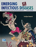
Tametomo no bui toukigami wo shirizoku zu [PDF - 147 KB - 1 page]
| EID | Potter P. Tametomo no bui toukigami wo shirizoku zu. Emerg Infect Dis. 2002;8(7):752. https://doi.org/10.3201/eid0807.ac0807 |
|---|---|
| AMA | Potter P. Tametomo no bui toukigami wo shirizoku zu. Emerging Infectious Diseases. 2002;8(7):752. doi:10.3201/eid0807.ac0807. |
| APA | Potter, P. (2002). Tametomo no bui toukigami wo shirizoku zu. Emerging Infectious Diseases, 8(7), 752. https://doi.org/10.3201/eid0807.ac0807. |