Volume 18, Number 8—August 2012
Dispatch
Putative Novel Genotype of Avian Hepatitis E Virus, Hungary, 2010
Cite This Article
Citation for Media
Abstract
To explore the genetic diversity of avian hepatitis E virus strains, we characterized the near-complete genome of a strain detected in 2010 in Hungary, uncovering moderate genome sequence similarity with reference strains. Public health implications related to consumption of eggs or meat contaminated by avian hepatitis E virus, or to poultry handling, require thorough investigation.
Avian hepatitis E virus (aHEV) is a causative agent of big liver and spleen disease (BLSD) in chickens (1), which manifests itself, as its name indicates, by hepatosplenomegaly. The etiologic association has been demonstrated worldwide, including Australia, North America, Asia, and Europe (2,3). In Hungary, BLSD was observed during the 1980s, but association of the disease with hepatosplenomegaly was not made in Hungary until 2008 (4,5). Ivanics et al. (5) reported outbreaks of aHEV infection and increased deaths of chickens caused by hepatitis and splenitis and reduced egg production in chicken flocks in Hungary. Tissue samples from chickens collected during 2010 were positive for aHEV by reverse transcription PCR (RT-PCR) (6,7). Paired samples indicated seroconversion upon inoculation as measured by serologic assay, strongly suggesting an etiologic association between BLSD and aHEV infection in the affected chicken flocks (5).
The aHEV is characterized by a small, nonenveloped virion and a 6.6-kb, capped, poly-A tailed single-stranded RNA genome. The genome encodes 3 open reading frames (ORFs). ORF1 is a polyprotein encoding putative functional domains of methyltransferase, papain-like cystein protease, helicase, and RNA polymerase. The capsid protein is encoded by ORF2; ORF3 encodes a multifunctional cytoskeleton-associated protein linked to viral morphogenesis and pathogenesis (1).
aHEV is ≈50% similar at the genomic level to mammalian HEV genotypes; recent proposals classify aHEV into a different taxon, possibly a new genus, within Hepeviridae. GenBank now includes 5 full or nearly complete aHEV genome sequences that could be classified into 1 of the 3 recognized aHEV genotypes. These 5 prototype strains with full or nearly complete genome sequences include a genotype 1 strain from Australia, 2 genotype 2 strains from the United States, and 2 genotype 3 strains from Europe and the People’s Republic of China (6,8–10). Additional putative genotypes have been identified through analysis of an ≈130-nt fragment of the helicase domain, suggesting that diversity within aHEV is higher than recognized (11). Our primary objective was the genetic characterization of an aHEV strain from the 2010 outbreak in Hungary to increase understanding of the origin and evolution of these emerging pathogens.
Preliminary sequence analysis of the amplicons obtained by the diagnostic HEV RT-PCR from the 2010 BLSD outbreaks in Hungary (strains identified by HUN prefix) showed a moderate genetic relationship with reference aHEV sequences deposited in GenBank (5). One strain, HUN-16773–2010, found in a sample of liver tissue, was selected for further analysis. Viral RNA was extracted by using the SV96 total RNA nucleic acid extraction kit (Promega, Madison, WI, USA). Oligonucleotides were designed for our primer walking sequencing strategy on the basis of aHEV genome sequence data (Table). Genomic fragments were amplified by using a OneStep RT-PCR Kit (QIAGEN, Hilden, Germany) as described by the manufacturer. Amplicons were subsequently subjected to direct sequencing (Biomi Ltd., Gödöllő, Hungary).
The final assembly was 6,543 bp (GenBank accession no. JN997392), with negligible amounts of missing information at the 5′ (75 nt) and 3′ ends preceding the poly A tail (36 nt). Nucleotide and deduced amino acid sequences of HUN-16773–2010 aligned well with the other 5 prototype aHEV strains. The nearly complete coding sequence (1,515 aa) with a missing 17-aa region was determined for ORF1. Predicted functional domains (12) were identified. The sequence similarities (Megalign; DNASTAR Inc., Madison, WI, USA) of ORF1 in comparison with genotypes 1–3 aHEV strains were 81.0%–83.9% at the nucleotide level and 93.0%–95.4% at the amino acid level.
Unique amino acid changes in the ORF1 of HUN-16773–2010 were found in 16 positions compared with the European prototype aHEV strain (not shown) (6). ORF2 encodes the 606-aa capsid protein. Similarities to prototype aHEV strains were 83.9%–87.3% at the nucleotide level and 98.2%–99.3% at the amino acid level. We observed 2 unique amino acid substitutions in positions A53S and Q/M473T, the second residue located within antigenic region II (6). ORF3 encoded an 87-aa polypeptide. This region also shared high (>92%) nt and (>90%) aa similarities with genotype 1–3 aHEVs.
Figure A1
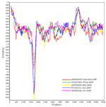
Figure A1. . . . Genomewide similarity of HUN-16773-2010 to 5 other avian hepatitis E virus (aHEV) strains with nearly complete genome sequences, 1 each belonging to genotypes 1–3. Sequences were analyzed with...
When analyzing the nearly complete genome sequences, we found that strain HUN-16773–2010 shared moderate similarity to genotypes 1–3 aHEV strains (nt, 81.9%–84.9%). Similar ranges of genome sequence similarity (nt, 81.8%–82.7%) distinguish prototype aHEV strains from each other. Using SimPlot analysis (http://sray.med.som.jhmi.edu/SCRoftware/simplot), we observed this moderate nucleotide similarity of HUN-16773–2010 to the 5 prototype strains along the entire genome (Figure A1), except the near-central region of ORF1, which showed less similarity among aHEV strains; this region encodes the hinge region flanking the papain-like protease and the X domain. Similarity in the genome was highest in the 5′ end of the genome and the overlapping region of ORF2 and ORF3. We found no evidence for recombination between HUN-16773–2010 and genotypes 1–3 strains (13), demonstrating that this strain was not generated through recombination from prototype aHEVs.
Figure
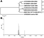
Maximum-likelihood and neighbor-joining trees were constructed with the substitution model of Tamura and Nei, including gamma distribution shape (G) parameter (TN93+G) as implemented in MEGA 5 (14); 500 bootstrap replicates were used to test the reliability of the tree topology. Phylogenetic analysis of the nearly complete aHEV genome revealed 4 major clusters, where genotypes 1–3 strains formed previously recognized branches (10); the aHEV strain, HUN-16773–2010, appeared to form a fourth cluster (Figure, panel A). To determine whether HUN-16773–2010 shared genetic relatedness in the partial helicase gene with recently identified variants of European aHEV strains (11), we reanalyzed this region by using a larger sample set. Our data reaffirmed that HUN-16773–2010 forms an independent branch (not shown).
To identify the evolutionary driving forces beyond the heterogeneity of aHEV genomes, we individually ran the alignments of all 3 ORFs on the DataMonkey server by using the random effects likelihood method (www.datamonkey.org). Evidence for strong selection pressure was found at the hinge region of ORF1 involving 17 sites between amino acid positions 556 and 605 (Figure, panel B). However, 905, 384, and 3 sites within ORF1, ORF2, and ORF3, respectively, were under purifying selection across aHEV genotypes.
Various genotypes of aHEV have been proposed to represent clusters of strains typical for particular geographic locations. However, new evidence has shown that some genotypes could be present on different continents (6,10,11). For example, although Australia seemingly remained the only continent with genotype 1 aHEV, different genotypes could be endemic to the Americas and particularly Europe, and these areas could show strain diversification. The origin of the novel aHEV strain in Hungary remains enigmatic; understanding its recent emergence awaits analysis of further isolates within and outside Europe.
Purifying selection was a key mechanism in the evolution of aHEV, suggesting the existence of a strong structural and functional constraint against diversification that would lead to extensive amino acid changes within the 3 ORFs in aHEV strains. In contrast, positive selection was predicted primarily in the hypervariable hinge region flanking the protease and X domains in ORF1. The biological role of this observation is not clear. However, recent findings have demonstrated that this region might be responsible for differences in replication efficiency (15). Thus, the positive evolutionary selection in this region could directly affect viral fitness and influence the pathogenesis of aHEV infections.
We provided further evidence for the marked sequence diversity among aHEVs by providing the nearly complete genome sequence of a strain identified during a BLSD outbreak in Hungary. Additional aHEV genomes could facilitate more penetrating insights into their epidemiologic features and evolutionary mechanisms and also could serve as molecular bases for reliable and robust demarcation criteria in future classification proposals.
Dr Bányai is the leader of Pathogen Discovery Group at Veterinary Medical Research Institute, Hungarian Academy of Sciences. His research interests include emerging and zoonotic diseases relevant to public health and veterinary medicine.
Acknowledgments
We thank Ottinger Ernőné and Ágnes Juhász for excellent technical assistance.
K.B. was supported by the “Lendület” Initiative of the Hungarian Academy of Sciences.
References
- Meng XJ. Hepatitis E virus: animal reservoirs and zoonotic risk. Vet Microbiol. 2010;140:256–65. DOIPubMedGoogle Scholar
- Payne CJ, Ellis TM, Plant SL, Gregory AR, Wilcox GE. Sequence data suggests big liver and spleen disease virus (BLSV) is genetically related to hepatitis E virus. Vet Microbiol. 1999;68:119–25. DOIPubMedGoogle Scholar
- Haqshenas G, Shivaprasad HL, Woolcock PR, Read DH, Meng XJ. Genetic identification and characterization of a novel virus related to human hepatitis E virus from chickens with hepatitis-splenomegaly syndrome in the United States. J Gen Virol. 2001;82:2449–62.PubMedGoogle Scholar
- Morrow CJ, Samu G, Mátrai E, Klausz A, Wood AM, Richter S, Avian hepatitis E virus infection and possible associated clinical disease in broiler breeder flocks in Hungary. Avian Pathol. 2008;37:527–35. DOIPubMedGoogle Scholar
- Ivanics É, Benyeda Z, Benyeda J, Nagy G, Dán Á, Tóth Á, Examination of hepatitis-E virus induced disease in Hungarian laying hen flocks [in Hungarian]. Magy Allatorv Lapja. 2011;133:587–93.
- Bilic I, Jaskulska B, Basic A, Morrow CJ, Hess M. Sequence analysis and comparison of avian hepatitis E viruses from Australia and Europe indicate the existence of different genotypes. J Gen Virol. 2009;90:863–73. DOIPubMedGoogle Scholar
- Johne R, Plenge-Bönig A, Hess M, Ulrich RG, Reetz J, Schielke A. Detection of a novel hepatitis E–like virus in faeces of wild rats using a nested broad-spectrum RT-PCR. J Gen Virol. 2010;91:750–8. DOIPubMedGoogle Scholar
- Billam P, Sun ZF, Meng XJ. Analysis of the complete genomic sequence of an apparently avirulent strain of avian hepatitis E virus (avian HEV) identified major genetic differences compared with the prototype pathogenic strain of avian HEV. J Gen Virol. 2007;88:1538–44. DOIPubMedGoogle Scholar
- Huang FF, Sun ZF, Emerson SU, Purcell RH, Shivaprasad HL, Pierson FW, Determination and analysis of the complete genomic sequence of avian hepatitis E virus (avian HEV) and attempts to infect rhesus monkeys with avian HEV. J Gen Virol. 2004;85:1609–18. DOIPubMedGoogle Scholar
- Zhao Q, Zhou EM, Dong SW, Qiu HK, Zhang L, Hu SB, Analysis of avian hepatitis E virus from chickens, China. Emerg Infect Dis. 2010;16:1469–72. DOIPubMedGoogle Scholar
- Marek A, Bilic I, Prokofieva I, Hess M. Phylogenetic analysis of avian hepatitis E virus samples from European and Australian chicken flocks supports the existence of a different genus within the Hepeviridae comprising at least three different genotypes. Vet Microbiol. 2010;145:54–61. DOIPubMedGoogle Scholar
- Koonin EV, Gorbalenya AE, Purdy MA, Rozanov MN, Reyes GR, Bradley DW. Computer-assisted assignment of functional domains in the nonstructural polyprotein of hepatitis E virus: delineation of an additional group of positive-strand RNA plant and animal viruses. Proc Natl Acad Sci U S A. 1992;89:8259–63. DOIPubMedGoogle Scholar
- Martin DP, Lemey P, Lott M, Moulton V, Posada D, Lefeuvre P. RDP3: a flexible and fast computer program for analyzing recombination. Bioinformatics. 2010;26:2462–3. DOIPubMedGoogle Scholar
- Tamura K, Peterson D, Peterson N, Stecher G, Nei M, Kumar S. MEGA5: Molecular Evolutionary Genetics Analysis using maximum likelihood, evolutionary distance, and maximum parsimony methods. Mol Biol Evol. 2011;28:2731–9. DOIPubMedGoogle Scholar
- Pudupakam RS, Kenney SP, Córdoba L, Huang YW, Dryman BA, Leroith T, Mutational analysis of the hypervariable region of hepatitis E virus reveals its involvement in the efficiency of viral RNA replication. J Virol. 2011;85:10031–40. DOIPubMedGoogle Scholar
Figures
Table
Cite This ArticleTable of Contents – Volume 18, Number 8—August 2012
| EID Search Options |
|---|
|
|
|
|
|
|
Please use the form below to submit correspondence to the authors or contact them at the following address:
Ádám Dán, Central Agricultural Office, Veterinary Diagnostic Directorate, Tábornok u. 2., H-1149, Budapest, Hungary
Top