Volume 18, Number 8—August 2012
Dispatch
New Variants of Porcine Epidemic Diarrhea Virus, China, 2011
Abstract
In 2011, porcine epidemic diarrhea virus (PEDV) infection rates rose substantially in vaccinated swine herds. To determine the distribution profile of PEDV outbreak strains, we sequenced the full-length spike gene from samples from 9 farms where animals exhibited severe diarrhea and mortality rates were high. Three new PEDV variants were identified.
A member of the family Coronaviridae, genus alphacoronavirus, porcine epidemic diarrhea virus (PEDV) is an enveloped, single-stranded positive-sense RNA virus (1). PEDV is the major causative agent of porcine epidemic diarrhea, which is characterized by severe enteritis, vomiting, watery diarrhea, and weight loss. PEDV infections have a substantial detrimental effect on the swine industry because the mortality rates are high, especially in sucking piglets (1). The major structural gene of the 28-kb PEDV genome encodes the multifunctional virulence factor, spike (S), which is responsible for viral receptor binding, induction of neutralizing antibodies, and host cell fusion. The S gene sequences are a distinguishing feature of PEDV strains, which affect virulence and evolution (2–4).
The first confirmed PED case in the People’s Republic of China was reported in 1973. Almost 2 decades later, an oil emulsion, inactivated vaccine was developed and has since been in wide use throughout the swine industry in China. Until 2010, the prevalence of PEDV infection was relatively low with only sporadic outbreaks; however, starting in late 2010, a remarkable increase in PED outbreaks occurred in the pig-producing provinces. The affected pigs exhibited watery diarrhea (Figure 1, panels A, B), dehydration with milk curd vomitus (Figure 1, panel C), and thin-walled intestines (Figure 1, panel D) with severe villus atrophy and congestion (Figure 1, panels E, F). The disease progressed to death within a few days. Pigs of all ages were affected and exhibited diarrhea and loss of appetite with different degrees of severity, which were determined to be age dependent; 100% of suckling piglets became ill. Pigs >2 weeks of age experienced mild diarrhea and anorexia, which completely resolved within a few days (5). Morbidity and mortality rates were lower for vaccinated herds than for nonvaccinated herds, which suggests the emergence of a new PEDV field strain(s) for which the current vaccine, based on the CV777 strain, was partially protective. To identify the PEDV strain(s) responsible for the recent outbreak in China, we sequenced the full-length S gene of isolates obtained from diarrhea samples collected from pigs at 9 affected pig farms.
From January 2011 through October 2011, a total of 455 samples (fecal, intestine, and milk) were collected from 57 farms in 12 provinces of China. All samples were evaluated by reverse transcription PCR (RT-PCR), by using previously described primers (6). Forty-five (78.95%) of the farms had at least 1 PEDV-positive sample. A total of 278 (61.11%) samples were PEDV positive, including 253 (of 402; 62.94%) fecal samples, 20 (of 31; 64.52%) intestine samples, and 5 (of 22; 22.73%) milk samples. The representative detection of PEDV in fecal samples of PED-affected farms is shown in Technical Appendix Figure 1.
Nine diarrhea samples were collected from pigs at 9 farms (where animals had severe diarrhea and mortality rate was high) for sequencing analysis of the full-length S gene (Technical Appendix Table 1). RT-PCR gene-specific primers were designed on the basis of the sequence of PEDV-CV777 strain (GenBank accession no. AF353511.1) (Table 1) and used to amplify 3 overlapping cDNA fragments spanning the entire S gene. The amplicons were sequenced in both directions (GenScript Co., Nanjing, PRC).
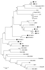
The 9 PEDV S gene sequences were aligned with the sequences of 24 previously published PEDV S genes (Table 2) by using the ClustalX (version 1.82), Bioedit (version 7.0.9.0) and MegAlign version 5.0 (DNAStar Inc., Madison, WI, USA) software packages (14). The full-length S gene sequences of the 9 isolates from our study showed overall high conservation with the reference strains, up to 94.9%–99.6% homology (Technical Appendix Table 2). By phylogenetic analysis, 4 of the field isolates (CH2, CH5, CH6, CH7) clustered with the previously described strain JS-2004–2 from China. Three field isolates (CH1, CH8, CHGD-01) formed a unique cluster with the sequence-confirmed variant strain CH-FJND-3, which had been isolated from China in 2011 (7). CH1 and CH8 were isolated from 2 farms, where all sucking piglets had died from diarrhea, even though all of the sows had been vaccinated with the PEDV-CV777 strain–based inactivated vaccine. The isolated variant strains, CHGD-01 and CH1, were tested in experimental infection studies and found to cause illness in 100% of sucking piglets (data not shown).
The phylogenetic analysis of the S gene nucleotide sequences revealed 3 major clusters (Figure 2). Clade 1 comprised 6 strains from our study (CH2, CH3, CH4, CH5, CH6, CH7), the vaccine strain CV777 from China, the attenuated strain DR13 from South Korea, and 2 strains (CHFJND-1, CHFJND-2) that had been isolated in China in 2011. Clade 2 consisted of 4 variant strains (CH1, CH8, CHFJND-3, CHGD-01) that were identified from China in 2011. Clade 3 was composed of 9 isolates from South Korea and 2 strains from Japan (NK and Kawahira). The deduced amino acids of the 4 variant strains in clade 2 had 93% homology to CV777. Furthermore, the 4 variant strains from China (CH1, CH8, CHGD-01, CH-FJND-3) and 9 PEDV isolates from South Korea shared a 5-aa insertion (at positions 56–60 of the S protein) with CV777. One amino acid insertion at position 141 was shared among all variant strains and 6 isolates from South Korea (Technical Appendix Figure 2). In the S genes, 132 point mutations were found that accounted for genetic diversity among the isolates.
The recent 4 isolates from China (CH2, CH5, CH6, CH7) were closely related to the previously identified isolates from China (JS-2004–2, LJB03, DX) and another 4 variant strains. Three of the new isolates (CH1, CH8, CHGD-01) were highly pathogenic in piglets. All strains were obtained from farms that used the CV777-based inactivated vaccine but had 100% prevalence of diarrhea in pigs (Technical Appendix Table 1). Another 2 field isolates (CH3, CH4) from 2 farms with pigs with severe diarrhea shared the highest sequence identity with attenuated strain DR13 from South Korea (99.2% and 99.1%, respectively), which has been in routine use as an oral vaccine against PEDV in South Korea since 2004 (15). The appearance of strains in China similar to those from South Korea and their role in the recent PEDV outbreak should be further investigated.
RT-PCR amplification and sequencing analysis of the full-length PEDV spike genes were used to investigate isolates from diarrhea samples from local pig farms with severe diarrhea in piglets. Both classical and variant strains were detected, implying a diverse distribution profile for PEDV on pig farms in China. The sequence insertions and mutations found in the variant strains may have imparted a stronger pathogenicity to the new PEDV variants that influenced the effectiveness of the CV777-based vaccine, ultimately causing the 2011 outbreak of severe diarrhea on China’s pig farms. Future studies should investigate the biologic role of these particular insertions and mutations. Furthermore, our study of the full-length S gene revealed a more comprehensive distribution profile that reflects the current PEDV status in pig farms in China, including the presence of a strain similar to strain DR13, isolated in South Korea. Collectively, these data indicate the urgent need to develop novel variant strain–based vaccines to treat the current outbreak in China.
Mr Li is a PhD student at Huazhong Agricultural University. His research interests are focused on animal pathogen isolation and pathogenic mechanisms.
Acknowledgment
This work was supported by grants from the National Swine Industry Research System (no. CARS-36) and the Special Project from Guangdong Science and Technology Department (no. 2010B090301020).
References
- Pensaert MB, De Bouck P. A new coronavirus-like particle associated with diarrhea in swine. Arch Virol. 1978;58:243–7. DOIPubMedGoogle Scholar
- Lee DK, Park CK, Kim SH, Lee C. Heterogeneity in spike protein genes of porcine epidemic diarrhea viruses isolated in Korea. Virus Res. 2010;149:175–82. DOIPubMedGoogle Scholar
- Sato T, Takeyama N, Katsumata A, Tuchiya K, Kodama T, Kusanagi K. Mutations in the spike gene of porcine epidemic diarrhea virus associated with growth adaptation in vitro and attenuation of virulence in vivo. Virus Genes. 2011;43:72–8. DOIPubMedGoogle Scholar
- Lee DK, Cha SY, Lee C. The N-terminal region of the porcine epidemic diarrhea virus spike protein is important for the receptor binding. Korean Journal of Microbiology and Biotechnology. 2011;39:140–5.
- Shibata I, Tsudab T, Moria M, Onoa M, Sueyoshib M, Urunoa K. Isolation of porcine epidemic diarrhea virus in porcine cell cultures and experimental infection of pigs of different ages. Vet Microbiol. 2000;72:173–82. DOIPubMedGoogle Scholar
- Zhang K, He Q. Establishment and clinical application of a multiplex reverse transcription PCR for detection of porcine epidemic diarrhea virus, porcine transmissible gastroenteritis virus and porcine group A rotavirus. Chinese Journal of Animal and Veterinary Sciences. 2010;41:1001–5.
- Chen J, Liu X, Shi D, Shi H, Zhang X, Feng L. Complete genome sequence of a porcine epidemic diarrhea virus variant. J Virol. 2012;86:3408. DOIPubMedGoogle Scholar
- Chen J, Wang C, Shi H, Qiu HJ, Liu S, Shi D, Complete genome sequence of a Chinese virulent porcine epidemic diarrhea virus strain. J Virol. 2011;85:11538–9. DOIPubMedGoogle Scholar
- Yeo SG, Hernandez M, Krell PJ, Nagy ÉÉ. Cloning and sequence analysis of the spike gene of porcine epidemic diarrhea virus Chinju99. Virus Genes. 2003;26:239–46. DOIPubMedGoogle Scholar
- Kang TJ, Seo JE, Kim DH, Kim TG, Jang YS, Yang MS. Cloning and sequence analysis of the Korean strain of spike gene of porcine epidemic diarrhea virus and expression of its neutralizing epitope in plants. Protein Expr Purif. 2005;41:378–83. DOIPubMedGoogle Scholar
- Park SJ, Song DS, Ha GW, Park BK. Cloning and further sequence analysis of the spike gene of attenuated porcine epidemic diarrhea virus DR13. Virus Genes. 2007;35:55–64. DOIPubMedGoogle Scholar
- Duarte M, Laude H. Sequence of the spike protein of the porcine epidemic diarrhoea virus. J Gen Virol. 1994;75:1195–200. DOIPubMedGoogle Scholar
- Kocherhans R, Bridgen A, Ackermann M, Tobler K. Completion of the porcine epidemic diarrhoea coronavirus (PEDV) genome sequence. Virus Genes. 2001;23:137–44. DOIPubMedGoogle Scholar
- Tamura K, Dudley J, Nei M, Kumar S. MEGA4: Molecular Evolutionary Genetics Analysis (MEGA) software version 4.0. Mol Biol Evol. 2007;24:1596–9. DOIPubMedGoogle Scholar
- Song D, Park B. Porcine epidemic diarrhoea virus: a comprehensive review of molecular epidemiology, diagnosis, and vaccines. Virus Genes. 2012;44:167–75. DOIPubMedGoogle Scholar
Figures
Tables
Cite This ArticleTable of Contents – Volume 18, Number 8—August 2012
| EID Search Options |
|---|
|
|
|
|
|
|


Please use the form below to submit correspondence to the authors or contact them at the following address:
Qigai He, State Key Laboratory of Agricultural Microbiology, College of Veterinary Medicine, Huazhong Agricultural University, Wuhan 430070, PRC
Top