Volume 6, Number 2—April 2000
Research
Serologic Response to Culture Filtrate Antigens of Mycobacterium ulcerans during Buruli Ulcer Disease
Figure
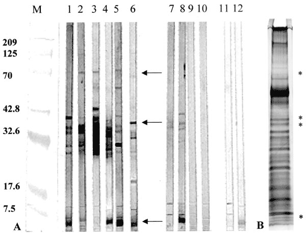
Figure. Western blot reactivity to and Silver stain analysis of Mycobacterium ulcerans culture filtrates (MUCF). A) Representative antibody responses to MUCF. M; molecular weight markers with annotations corresponding to molecular weight on the left; lanes 1-6, representative BU patient sera with reactivity to MUCF; lanes 7-10, representative antibody reactivity in healthy persons from the disease-endemic area; lanes 11-12, serologic reactivities to MUCF of two representative tuberculosis (TB) patients. TB patient sera that were serologically reactive to MUCF were included as a control for cross-reactivity. Regions of antibody reactivity corresponding to M. ulcerans antigens of 70, 38/36, and 5 kDa are noted (arrows). B) Silver-stained SDS-PAGE gel of MUCF. The identification of putative proteins corresponding to the serologically reactive 70, 38/36, and 5 kDa MUCF antigens are noted (stars). For Western blot analyses, aliquots (50 µg protein) of MUCF were resolved by discontinuous SDS-PAGE (10) with preparative 10% to 20% gradient 1-well gels (Novex, San Diego, CA) and then transferred to nitrocellulose (14). Nitrocellulose sheets were cut into 2-mm strips and stored in 5% skim milk in Tris-buffered saline, pH 7.6, until used. Antibody to MUCF was detected by probing nitrocellulose strips with sera at a 1:50 (vol:vol dilution). Bound serum antibodies were detected with alkaline phosphatase-conjugated anti-human antibody (Sigma Chemical Co., St. Louis, MO), and the substrate 4-Bromo-3-Chloro-2-Indoyl-1-Phosphate/Nitro blue tetrazoleum (Sigma Chemical Co., St. Louis, MO). All sera were tested in duplicate for confirmation of the antibody responses.
References
- Horsburgh CR Jr, Meyers WM. Buruli ulcer. In: Pathology of emerging infections. Horsburgh CR Jr, Nelson AM, editors. Washington: American Society for Mibrobiology Press; 1997. p. 119-26.
- Dobos KM, Horsburgh CR Jr, Ashford DA, Quinn FD, King CH. Emergence of a unique group of necrotizing mycobacterial diseases. Emerg Infect Dis. 1999;5:367–78. DOIPubMedGoogle Scholar
- Marston BJ, Diallo MO, Horsburgh CR Jr, Diomande I, Saki MZ, Kanga J-M, Emergence of Buruli ulcer disease in the Daloa region of Côte d'Ivoire. Am J Trop Med Hyg. 1995;52:219–24.PubMedGoogle Scholar
- Amofah GK, Sagoe-Moses C, Adjei-Acquah C, Frimpong EH. Epidemiology of Buruli ulcer in Amansie West district, Ghana. Trans R Soc Trop Med Hyg. 1993;87:644–5. DOIPubMedGoogle Scholar
- Hayman J. Postulated epidemiology of Mycobacterium ulcerans infection. Int J Epidemiol. 1991;20:1093–8. DOIPubMedGoogle Scholar
- Portaels F, Elsen P, Guimaraes-Peres A, Fonteyne PA, Meyers WM. Insects in the transmission of Mycobacterium ulcerans infection. Lancet. 1999;353:986. DOIPubMedGoogle Scholar
- Asiedu K, Etuaful S. Socioeconomic implications of Buruli ulcer in Ghana: a three-year review. Am J Trop Med Hyg. 1998;59:1015–22.PubMedGoogle Scholar
- Amofah G, Asamoah S, Afram-Gyening G. Effectiveness of excision of pre-ulcerative Buruli lesions in field situations in a rual district in Ghana. Trop Doct. 1998;28:81–3.PubMedGoogle Scholar
- Stanford TL, Revill WDL, Gunthorpe WJ, Grange JM. The production and preliminary investigation of Burulin, a new skin test for Mycobacterium ulcerans infection. J Hyg (Lond). 1975;74:7–16. DOIPubMedGoogle Scholar
- Laemmli UK. Cleavage of structural proteins during the assembly of the head of bacteriophage T4. Nature. 1970;227:680–5. DOIPubMedGoogle Scholar
- Morrissey JH. Silver stain for proteins in polyacrylamide gels: a modified procedure with enhanced, uniform sensitivity. Anal Biochem. 1981;117:307–10. DOIPubMedGoogle Scholar
- O'Farrell PH. High resolution two dimensional analysis of proteins. J Biol Chem. 1975;250:4007–21.PubMedGoogle Scholar
- Sonnenberg MG, Belisle JT. Definition of Mycobacterium tuberculosis culture filtrate proteins by two dimensional polyacrylamide gel electrophoresis, N-terminal amino acid sequencing, and electrospray mass spectrometry. Infect Immun. 1997;65:4515–24.PubMedGoogle Scholar
- Towbin H, Staehelin T, Gordon J. Electrophoretic transfer of protein from polyacrylamide gels to nitrocellulose sheets: procedure and some applications. Proc Natl Acad Sci U S A. 1979;76:4350–4. DOIPubMedGoogle Scholar
- Dobos KM, Khoo KH, Swiderek KM, Brennan PJ, Belisle JT. Definition of the full extent of glycosylation of the 45-kilodalton glycoprotein of Mycobacterium tuberculosis. J Bacteriol. 1996;178:2498–506.PubMedGoogle Scholar
- De Vries RR. An immunogenetic view of delayed type hypersensitivity. Tubercle. 1991;72:161–7. DOIPubMedGoogle Scholar
- Sengupta U. Studies on lepromin and soluble antigens of M. leprae: their classification standardization and use. Indian J Lepr. 1991;63:457–65.PubMedGoogle Scholar
- Shield MJ, Stanford JL, Paul RC, Carswell JW. Multiple skin testing of tuberculosis patients with a range of new tuberculins, and a comparison with leprosy and Mycobacterium ulcerans infection. J Hyg (Lond). 1977;78:331–48. DOIPubMedGoogle Scholar
- Dannenberg AM Jr. Delayed-type hypersensitivity and cell-mediated immunity in the pathogenesis of tuberculosis. Immunol Today. 1991;7:228–33. DOIGoogle Scholar
- Orme IM, Andersen P, Boom WH. T cell response to Mycobacterium tuberculosis. J Infect Dis. 1993;167:1481–97.PubMedGoogle Scholar
- Belisle JT, Vissa VD, Sievert T, Takayama K, Brennan PJ, Besra GS. Role of the major antigen of Mycobacterium tuberculosis in cell wall biogenesis. Science. 1997;276:1420–2. DOIPubMedGoogle Scholar