Volume 20, Number 5—May 2014
Dispatch
Francisella tularensis subsp. tularensis Group A.I, United States
Cite This Article
Citation for Media
Abstract
We used whole-genome analysis and subsequent characterization of geographically diverse strains using new genetic signatures to identify distinct subgroups within Francisella tularensis subsp. tularensis group A.I: A.I.3, A.I.8, and A.I.12. These subgroups exhibit complex phylogeographic patterns within North America. The widest distribution was observed for A.I.12, which suggests an adaptive advantage.
Tularemia, caused by the bacterium Francisella tularensis, is a potentially severe disease that often causes unspecific symptoms; because of its low infectious dose and ease of dissemination, F. tularensis is considered a category A biothreat agent (1). Three subspecies of F. tularensis have been identified; F. tularensis subsp. tularensis (type A) has been identified only in North America. Numerous subtyping schemes have subdivided type A into 2 groups, A.I and A.II (2–8). Group A.II is found primarily in the western United States (3,4), whereas group A.I is found throughout the central and eastern regions of the country and sporadically in some western states (3,4,9).
Groups A.I and A.II differ in virulence, as do subgroups within A.I, although clinical signs and symptoms can be similar. Human infections involving A.I strains are associated with a higher fatality rate than that for infections involving A.II strains (4,10); this finding was experimentally confirmed in mice (11). Kugeler et al. (10) used pulsed-field gel electrophoresis (PFGE) to identify 2 subgroups within A.I, A1a and A1b; this study found A1b strains were associated with higher death rates and were more often isolated from human tissue types that were associated with severe disease. This difference was also experimentally confirmed in mice (11,12). However, virulence testing is not often used in clinical settings because it is slow, complicated, and expensive. Thus, molecular approaches that can rapidly assign an unknown strain to one of the recognized groups with known differences in virulence may provide valuable information to clinicians.
Figure 1
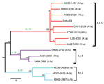
Figure 1. Neighbor-joining tree of 14 Francisella tularensis subsptularensis group A.I strains constructed on the basis of single-nucleotide polymorphisms (SNPs) discovered from whole-genome sequencingLines represent major groups within A.I: red, A.I.12; purple, A.I.8;...
Because PFGE lacks the phylogenetic resolution of some other testing methods (6), we independently identified genetic subgroups within A.I by conducting whole-genome sequencing (WGS) of 13 A.I strains (Figure 1; Table 1, Appendix). The 13 strains were selected on the basis of assignment to PFGE subgroups A1a or A1b (10) and to maximize geographic diversity; the previously sequenced A.I strain Schu S4 (13) was also included. WGS data were generated, assembled, and analyzed as described in the Technical Appendix (wwwnc.cdc.gov/EID/article/20/5/13-1559-Techapp1.pdf).
Our whole-genome phylogeny revealed 3 major subgroups within F. tularensis subsp. tularensis A.I: A.I.3, A.I.8, and A.I.12 (Figure 1). The names we assigned to these subgroups are consistent with previous phylogenetic nomenclature within F. tularensis (14). With the exception of 1 strain (ND01-1900) that was not assigned to any of the 3 subgroups, all strains previously assigned to PFGE subgroup A1a belonged to the newly designated A.I.12 subgroup (Figure 1; Table 1). In contrast, strains previously assigned to PFGE subgroup A1b were distributed among all 3 of the new subgroups (Figure 1; Table 1). We concluded that results of characterization of subgroups A1a and A1b by PFGE are not in agreement with findings of a robust whole-genome phylogeny and therefore focused the remainder of our analysis on subgroups identified by using WGS.
We observed several differences among the 3 subgroups in the whole-genome phylogeny (Figure 1). The first split separated the A.I.3 subgroup from the A.I.8 and A.I.12 subgroups; a second split separated the A.I.8 and A.I.12 subgroups. A long branch of 25 single nucleotide polymorphisms (SNPs) led to the A.I.3 subgroup, in which relatedness among the sequenced strains was moderate. A branch of 9 SNPs led to the A.I.8 subgroup, and again, relatedness among the sequenced strains was moderate. The branch leading to subgroup A.I.12 was, by comparison, much longer (37 SNPs), and the sequenced strains were separated only by 3 short branches (1–4 SNPs). This pattern of several short branches without hierarchical structuring is consistent with a recent radiation, an evolutionary process in response to adaptive change, new ecologic opportunities, or a combination of these factors.
Figure 2
Figure 2. Geographic alignment of 179 geographically diverse Francisella tularensis subsptularensis A.I strains, by subgroup, United StatesA) Canonical single-nucleotide polymorphism (canSNP) topology of 15 intervening and terminal subpopulations defined by screening of 16...
To show more comprehensive phylogenetic patterns, we developed 16 canonical SNP (canSNP) assays as described (Technical Appendix) and used them to screen 179 F. tularensis subsp. tularensis A.I strains selected from the collections of the Centers for Disease Control and Prevention (Fort Collins, CO, USA). We selected strains that were representative of all states where A.I infections occur and of all PFGE classification types (Table 1). One limitation of our study is that we did not analyze an equal number of strains from all regions of the country. However, our sample reflects the distribution of human disease caused by F. tularensis subsp. tularensis A.I strains: prevalent in the central United States, less common in the eastern United States, and rare in the western United States (4). The canSNP assays were based on 12 SNP signatures (Table 2) from the whole-genome phylogeny (Figure 1) and 4 previously described SNP signatures (6–8). Using these assays, we assigned the 179 strains to 15 F. tularensis subsp. tularensis A.I subpopulations, including 8 intervening nodes (Figure 2, panel A). We found 6 subpopulations in the A.I.12 subgroup, 4 in A.I.8, and 4 in A.I.3 (Table 1). To identify broad phylogeographic patterns, we created maps indicating specific states where strains from the 15 subpopulations were isolated (Figure 2, panel B). Within these maps, we created boundaries corresponding to 3 regions within the United States: western, central, and eastern.
Each subgroup exhibited complex yet distinct phylogeographic patterns (Figure 2, panel B). Group A.I.12 strains, assigned to 6 subpopulations (Figure 2, panel A), were isolated throughout the United States: all 6 subpopulations were found in the central region, 3 in the western region, and 5 in the eastern region (Figure 2, panel B, top). Group A.I.8 strains, assigned to 4 subpopulations, were found in the central (3 subpopulations) and western (including Alaska and British Columbia; 3 subpopulations) regions, but only 1 strain was isolated in the eastern region (Figure 2, panel B, middle). For group A.I.3 strains, assigned to 4 subpopulations, distribution differed dramatically from the other subgroups; most strains and all 4 subpopulations occurred in the eastern region and just 1 subpopulation in the central region but none in the western region (Figure 2, panel B, bottom).
The occurrence of the A.I.3 subgroup in the eastern United States could be a recent or ancient event. The subgroup may have been introduced more recently from the central region to a naive niche in the eastern region through importation of rabbits (Sylvilagus floridanus) as recently as the 1920s (3); before 1937, tularemia was nearly nonexistent in the eastern region (15). If the introduction is recent, the current lack of A.I.3 strains in the central United States could be the result of a selective sweep that nearly eliminated this subgroup from its geographic origin. However, most strains and genetic diversity (i.e., subpopulations) within the A.I.3 subgroup are found in the eastern United States, which may reflect a more ancient history in this region involving early introduction and establishment of this subgroup east of the Appalachian Mountains, with only recent spread to the central region.
If we assume that the greatest genetic diversity in a phylogenetic context implies ancient origins, our findings suggest that the central United States is the likely geographic origin of a common ancestor to F. tularensis subsp. tularensis subgroups A.I.12 and A.I.8 and, perhaps, the A.I group as a whole. The large geographic range of the A.I.12 subgroup and the phylogenetic pattern of a long branch leading to a polytomy with genetic homogeneity point to a possible adaptive advantage for this subgroup. This advantage may be related to difference in virulence among A.I strains, as suggested by previous testing in mice of 2 A.I.12 strains that exhibited lower virulence than that of 2 A.I.3 strains (11). Further research is needed to determine whether the genomic differences that define this subgroup are associated with known F. tularensis virulence determinants.
Dr Birdsell is a postdoctoral fellow at the Center for Microbial Genetics and Genomics, Northern Arizona University. Her primary research interest is the evolution of F. tularensis.
Acknowledgments
We thank Mia Champion for her assistance with initial analyses and Laurel Respicio-Kingry for her assistance with SNP genotyping.
This work was supported in part by the US Department of Homeland Security Science and Technology Directorate through award HSHQDC-10-C-00139, the Swedish Civil Contingencies Agency through TA#014-2010-01, the Laboratory for Molecular Infection Medicine Sweden , and Västerbotten County Council. M.G. was supported by the Lendület program of the Hungarian Academy of Sciences.
References
- Rotz LD, Khan AS, Lillibridge SR, Ostroff SM, Hughes JM. Public health assessment of potential biological terrorism agents. Emerg Infect Dis. 2002;8:225–30 . DOIPubMedGoogle Scholar
- Johansson A, Farlow J, Larsson P, Dukerich M, Chambers E, Byström M, Worldwide genetic relationships among Francisella tularensis isolates determined by multiple-locus variable-number tandem repeat analysis. J Bacteriol. 2004;186:5808–18 . DOIPubMedGoogle Scholar
- Farlow J, Wagner DM, Dukerich M, Stanley M, Chu M, Kubota K, Francisella tularensis in the United States. Emerg Infect Dis. 2005;11:1835–41 . DOIPubMedGoogle Scholar
- Staples JE, Kubota KA, Chalcraft LG, Mead PS, Petersen JM. Epidemiologic and molecular analysis of human tularemia, United States, 1964–2004. Emerg Infect Dis. 2006;12:1113–8. DOIPubMedGoogle Scholar
- Svensson K, Larsson P, Johansson D, Byström M, Forsman M, Johansson A. Evolution of subspecies of Francisella tularensis. J Bacteriol. 2005;187:3903–8. DOIPubMedGoogle Scholar
- Pandya GA, Holmes MH, Petersen JM, Pradhan S, Karamycheva SA, Wolcott MJ, Whole genome single nucleotide polymorphism based phylogeny of Francisella tularensis and its application to the development of a strain typing assay. BMC Microbiol. 2009;9:213 . DOIPubMedGoogle Scholar
- Svensson K, Granberg M, Karlsson L, Neubauerova V, Forsman M, Johansson A. A real-time PCR array for hierarchical identification of Francisella isolates. PLoS ONE. 2009;4:e8360 . DOIPubMedGoogle Scholar
- Vogler AJ, Birdsell D, Price LB, Bowers JR, Beckstrom-Sternberg SM, Auerbach RK, Phylogeography of Francisella tularensis: global expansion of a highly fit clone. J Bacteriol. 2009;191:2474–84. DOIPubMedGoogle Scholar
- Keim P, Johansson A, Wagner DM. Molecular epidemiology, evolution, and ecology of Francisella. Ann N Y Acad Sci. 2007;1105:30–66 . DOIPubMedGoogle Scholar
- Kugeler KJ, Mead PS, Janusz AM, Staples JE, Kubota KA, Chalcraft LG, Molecular epidemiology of Francisella tularensis in the United States. Clin Infect Dis. 2009;48:863–70. DOIPubMedGoogle Scholar
- Molins CR, Delorey MJ, Yockey BM, Young JW, Sheldon SW, Reese SM, Virulence differences among Francisella tularensis subsp. tularensis clades in mice. PLoS ONE. 2010;5:e10205. DOIPubMedGoogle Scholar
- Twine SM, Shen H, Kelly JF, Chen W, Sjostedt A, Conlan JW. Virulence comparison in mice of distinct isolates of type A Francisella tularensis. Microb Pathog. 2006;40:133–8. DOIPubMedGoogle Scholar
- Larsson P, Oyston PC, Chain P, Chu MC, Duffield M, Fuxelius HH, The complete genome sequence of Francisella tularensis, the causative agent of tularemia. Nat Genet. 2005;37:153–9. DOIPubMedGoogle Scholar
- Gyuranecz M, Birdsell DN, Splettstoesser W, Seibold E, Beckstrom-Sternberg SM, Makrai L, Phylogeography of Francisella tularensis subsp. holarctica, Europe. Emerg Infect Dis. 2012;18:290–3. DOIPubMedGoogle Scholar
- Ayres JC, Feemster R. Epidemiology of tularemia in Massachusetts with a review of the literature. N Engl J Med. 1948;238:187–94. DOIPubMedGoogle Scholar
Figures
Tables
Cite This ArticleTable of Contents – Volume 20, Number 5—May 2014
| EID Search Options |
|---|
|
|
|
|
|
|
Please use the form below to submit correspondence to the authors or contact them at the following address:
Corresponding author: David M. Wagner, Center for Microbial Genetics and Genomics, Northern Arizona University, Flagstaff, AZ 86011-4073, USACorresponding author: David M. Wagner, Center for Microbial Genetics and Genomics, Northern Arizona University, Flagstaff, AZ 86011-4073, USA
Top