Volume 11, Number 12—December 2005
Research
Echinococcosis in Tibetan Populations, Western Sichuan Province, China
Li Tiaoying*, Qiu Jiamin* , Yang Wen*, Philip S. Craig†, Chen Xingwang*, Xiao Ning*‡, Akira Ito‡, Patrick Giraudoux§, Mamuti Wulamu‡, Yu Wen*, and Peter M. Schantz¶
Figure 3
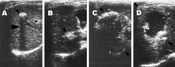
Figure 3. Lesions of alveolar echinococcosis (AE) by abdominal ultrasound examination. A) Calcified lesion: hyperechoic structure with a typical posterior shadow. B) Nodular hyperechoic lesion. C) Typical AE lesion: nonhomogeneous hyperechoic partially calcified area, without central necrosis. D) Typical AE lesion with central necrosis.
Page created: February 02, 2012
Page updated: February 02, 2012
Page reviewed: February 02, 2012
The conclusions, findings, and opinions expressed by authors contributing to this journal do not necessarily reflect the official position of the U.S. Department of Health and Human Services, the Public Health Service, the Centers for Disease Control and Prevention, or the authors' affiliated institutions. Use of trade names is for identification only and does not imply endorsement by any of the groups named above.