Volume 12, Number 11—November 2006
Dispatch
Recurrent Tuberculosis and Exogenous Reinfection, Shanghai, China
Figure 2

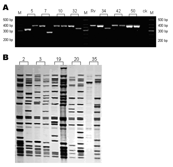
Figure 2. Genotyping analysis of clinical isolates from patients with recurrent tuberculosis. Numbers represented the patients' codes. A) Gel electrophoresis analysis of the PCR products of the mycobacterial interspersed repetitive unit (MIRU) locus 10. bp, base pair; M: DNA marker; Rv, H37Rv positive control; ck, negative control. B) IS6110 restriction fragment length polymorphism analysis of some patients with different MIRU patterns.
Page created: October 14, 2011
Page updated: October 14, 2011
Page reviewed: October 14, 2011
The conclusions, findings, and opinions expressed by authors contributing to this journal do not necessarily reflect the official position of the U.S. Department of Health and Human Services, the Public Health Service, the Centers for Disease Control and Prevention, or the authors' affiliated institutions. Use of trade names is for identification only and does not imply endorsement by any of the groups named above.