Volume 12, Number 3—March 2006
Letter
"Mycobacterium tilburgii" Infections
Cite This Article
Citation for Media
To the Editor: Advanced molecular biologic methods have improved the species differentiation and taxonomic classification of microorganisms, including nontuberculous mycobacteria. Identifying and characterizing an increasing number of "new" mycobacterial species of medical importance is now possible. Often, these newly described mycobacteria have been isolated from immunocompromised patients (1,2). Some of those have been difficult to cultivate, and 16S rRNA gene sequencing or similar methods have become of major importance to allow species identification and clinical diagnosis. Here, we report 2 patients with disease likely caused by a novel mycobacterial species that could not been previously cultivated. Diagnosis relied on molecular identification of acid-fast organisms in tissues. We also briefly review 2 similar cases published previously and note that all 4 known patients were from central Europe.
A 43-year-old woman without evidence of immunodeficiency reported recurrent episodes of dysuria, hematuria, and abdominal discomfort for >1 year. On cystoscopy, a hyperemic bladder with yellow plaques was observed. Biopsy of the plaques showed granulomatous infiltration of histiocytes. No definite diagnosis was made, and symptomatic relief occurred after a trial of empiric antimicrobial drug therapy. When the patient sought treatment again with persistent abdominal discomfort, endoscopy showed a lesion in the stomach that resembled a healed ulcer and numerous elevated yellow plaques throughout the colon and ileum. Microscopically, a granulomatous inflammation with macrophages filled with many acid-fast rods was seen, but mycobacterial growth did not occur in different media or in a guinea pig. Antituberculous treatment was initiated, and the patient slowly improved. A repeat colonoscopy showed fewer and smaller lesions. Efforts to culture the organism from biopsy specimens were again unsuccessful (different solid and liquid media, blood or chocolate agar, guinea pig, Balb/c mice). Sequencing of polymerase chain reaction (PCR) products of the 16S rRNA gene (3) from the organism represented a previously unknown mycobacterial species (EMBL DNA database: accession number Z50172). "Mycobacterium tilburgii" was proposed as the designation of this species because the novel mycobacterium was identified in the city of Tilburg (4). Retrospective analysis of the initial bladder biopsy specimen and of 2 lymph nodes taken during abdominal surgery (which became necessary because of a complicating ileal obstruction) confirmed the presence of "M. tilburgii" 16S rRNA gene sequences. All samples yielding PCR fragments hybridized with a "M. tilburgii"–specific biotinylated DNA probe.
Figure A1
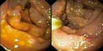
Figure A1. Yellow plaques in the mucosa of the duodenum in patient 2. Similar lesions had been observed in patient 1 in the colon, ileum, and bladder, whereas white lesion had been described...
A 34-year-old AIDS patient sought treatment for involuntary weight loss. Endoscopy showed white superficial embossments of the duodenal mucosa. Biopsy specimens were negative for acid-fast bacteria and mycobacterial growth (again, different solid and liquid media, extended incubation periods). Antiretroviral therapy was begun, but asthenia, persistent fever, diarrhea, vomiting, and cachexia developed. Repeat endoscopy showed yellow, plaquelike lesions in the duodenum (Figure A1) and esophagus with periodic acid–Schiff (PAS) and acid-fast intracellular bacteria that were nonculturable. PCR of 16S rRNA gene (3) confirmed the presence of mycobacterial DNA; sequencing showed 100% homology to the species with the proposed name "M. tilburgii" (4). Retrospectively, "M. tilburgii"16S rRNA was present in the initial duodenal biopsy specimen, when no mycobacteria were detected microscopically. Combination therapy led to a gradual disappearance of fever and diarrhea. The patient's weight increased, and the lesions disappeared (Table). Control endoscopy results were unremarkable, although duodenal biopsy specimens showed PAS-positive material. Mycobacterial DNA was no longer detectable.
We believe that the identified acid-fast organism named "M. tilburgii" was the causative agent of illness in the 2 patients. First, the clinical and histopathologic appearance of the lesions is compatible with mycobacterial infection. Second, the finding that the macrophages in these lesions contained large numbers of acid-fast bacteria supports the conclusion that mycobacterial infection caused the lesion. Third, the involvement of normally sterile locations such as lymph nodes, and the improvement after antimycobacterial therapy with disappearance of the lesions, acid-fast organisms, and mycobacterial DNA supports a causative role of the organism rather than a bystander role.
Attempts to culture the organism from biopsy specimens and resection materials remained unsuccessful, although specific requirements for organisms like M. genavense, a mycobacterium that also is not cultivable on regular mycobacterial media (5), were met. Diagnosis therefore had to rely on sequencing of PCR products. Based on 16S rRNA homology, "M. tilburgii" shows close relationship to M. sherrisii, a M. simiae–related mycobacterium (6) that probably corresponds to Mycobacterium strain IWGMT 90143 (accession no. X88906) (7). "M. tilburgii" also has a high homology to Mycobacterium sp. Murphy, a mycobacterial species that is considered the cause of canine lepra and that also has not been cultivated so far (8) (accession no. AF144747).
These 2 patients double the number of patients reported previously (Table). Both previously reported M. tilburgii patients had AIDS, 1 of whom had a disease similar to that of the second patient in this report (9). Taken together, 3 patients had gastrointestinal involvement with yellow mucosal plaques (Figure A1), and 1 patient had pulmonary nodules (10). One of the 4 patients did not have a detectable immunodeficiency, yet had gastrointestinal and urinary bladder lesions. Although complications occurred, treatment was successful in all 4 cases with usual anti–Mycobacterium avium complex therapy. Interestingly, all 4 patients so far are of middle-European descent. This species may be geographically confined, similar to the occurrence of M. malmoense in which most clinical isolates come from northern Europe (1).
References
- Brown-Elliott BA, Griffith DE, Wallace RJ Jr. Newly described or emerging human species of nontuberculous mycobacteria. Infect Dis Clin North Am. 2002;16:187–220. DOIPubMedGoogle Scholar
- Wagner D, Young LS. Nontuberculous mycobacterial infections. Infection. 2004;32:257–70. DOIPubMedGoogle Scholar
- Kirschner P, Springer B, Vogel U, Meier A, Wrede A, Kiekenbeck M, Genotypic identification of mycobacteria by nucleic acid sequence determination: report of a 2-year experience in a clinical laboratory. J Clin Microbiol. 1993;31:2882–9.PubMedGoogle Scholar
- Buiting A, Vos M, Bergmans A, Schouls L. A new mycobacterial species causing disseminated infection. 35th Interscience Conference on Antimicrobial Agents and Chemotherapy. San Francisco 1995; Abstract K45. Washington: American Society for Microbiology; 1995.
- Realini L, De RK, Hirschel B, Portaels F. Blood and charcoal added to acidified agar media promote the growth of Mycobacterium genavense. Diagn Microbiol Infect Dis. 1999;34:45–50. DOIPubMedGoogle Scholar
- Selvarangan R, Wu WK, Nguyen TT, Carlson LD, Wallis CK, Stiglich SK, Characterization of a novel group of mycobacteria and proposal of Mycobacterium sherrisii sp. nov. J Clin Microbiol. 2004;42:52–9. DOIPubMedGoogle Scholar
- Wayne LG, Good RC, Bottger EC, Butler R, Dorsch M, Ezaki T, Semantide- and chemotaxonomy-based analyses of some problematic phenotypic clusters of slowly growing mycobacteria, a cooperative study of the International Working Group on Mycobacterial Taxonomy. Int J Syst Bacteriol. 1996;46:280–97. DOIPubMedGoogle Scholar
- Hughes MS, James G, Ball N, Scally M, Malik R, Wigney DI, Identification by 16S rRNA gene analyses of a potential novel mycobacterial species as an etiological agent of canine leproid granuloma syndrome. J Clin Microbiol. 2000;38:953–9.PubMedGoogle Scholar
- Richter E, Rusch-Gerdes S, Niemann S, Stoehr A, Plettenberg A. Detection, identification, and treatment of a novel, non-cultivable Mycobacterium species in an HIV patient. AIDS. 2000;14:1667–8. DOIPubMedGoogle Scholar
- Kolditz M, Halank M, Spornraft-Ragaller P, Schmidt H, Höffken G. Localized pulmonary infection associated with Mycobacterium tilburgii in an HIV-infected patient. Infection. 2005;33:278–81. DOIPubMedGoogle Scholar
Figure
Table
Cite This ArticleRelated Links
Table of Contents – Volume 12, Number 3—March 2006
| EID Search Options |
|---|
|
|
|
|
|
|
Please use the form below to submit correspondence to the authors or contact them at the following address:
Dirk Wagner, Division of Infectious Diseases, University Hospital, Hugstetter Str 55, D-79106 Freiburg, Germany; fax: 49-761-270-820
Top