Volume 12, Number 6—June 2006
Letter
Linguatuliasis in Germany
Figure

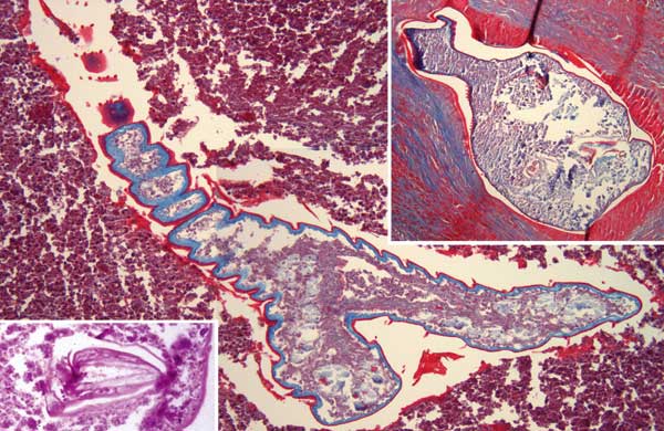
Figure. Linguatula serrata nymphs in lung tissue. Main panel shows the parasite's serrated nature and the cuticular spines (magnification ×200, Masson trichrome stain). Right upper inset, pulmonary nodule with prominent fibrotic reaction and shed cuticle around 1 nymph (magnification ×200, Masson trichrome stain). Left lower inset, detailed view of 1 parasite hook (magnification ×630, hematoxylin and eosin stain).
Page created: January 04, 2012
Page updated: January 04, 2012
Page reviewed: January 04, 2012
The conclusions, findings, and opinions expressed by authors contributing to this journal do not necessarily reflect the official position of the U.S. Department of Health and Human Services, the Public Health Service, the Centers for Disease Control and Prevention, or the authors' affiliated institutions. Use of trade names is for identification only and does not imply endorsement by any of the groups named above.