Volume 16, Number 5—May 2010
Dispatch
Streptococcus dysgalactiae subsp. equisimilis Bacteremia, Finland, 1995–2004
Cite This Article
Citation for Media
Abstract
We conducted a retrospective population-based study of 140 episodes of Streptococcus dysgalactiae subsp. equisimilis bacteremia occurring in Finland during 1995–2004. Rare emm types were associated with more severe disease and increased mortality rates. Skin and soft tissue infections were more frequent clinical signs among cases caused by common emm types.
Lancefield groups C and G β-hemolytic streptococci (GCS and GGS) may colonize the pharynx, skin, gastrointestinal tract, and female genitourinary tracts (1). According to recent taxonomic studies, large colony-forming groups C and G streptococci that infect humans are classified as Streptococcus dysgalactiae subsp. equisimilis (2). S. dysgalactiae subsp. equisimilis and S. pyogenes share virulence factors (3,4). The M protein is an important virulence factor because it confers resistance to phagocytosis (5). As with emm genes of S. pyogenes, the emm homologs of groups C and G S. dysgalactiae subsp. equisimilis are used for sequence-based typing (4,6,7), with >50 sequence types currently described (www.cdc.gov/ncidod/ biotech/strep/emmtypes.htm). The aim of our study was to determine the clinical signs, epidemiologic characteristics, and emm types of S. dysgalactiae subsp. equisimilis bacteremia during the 10-year observation period in Finland.
We retrospectively reviewed the medical records of all adult patients (>16 years of age) in Pirkanmaa Health District, Finland, with >1 blood cultures positive for group C or group G S. dysgalactiae subsp. equisimilis from January 1995 through December 2004. The Pirkanmaa Health District (460,000 inhabitants) has 1 tertiary care hospital (Tampere University Hospital) and 4 other hospitals (Hatanpää City Hospital and the District Hospitals in Valkeakoski, Vammala, and Mänttä). Laboratory records were screened to identify all blood cultures positive for group C or group G S. dysgalactiae subsp. equisimilis during the study period. Our case definition included all patients who had a positive blood culture for S. dysgalactiae subsp. equisimilis and clinical signs compatible with septicemia. A severe disease was defined as a septicemia leading to death or needing intensive care unit treatment. All 128 GGS isolates and 12 of 18 GCS isolates were confirmed to be S. dysgalactiae subsp. equisimilis. Thus, these 140 episodes of S. dysgalactiae subsp. equisimilis septicemia (involving 137 patients) comprised the present study. Two of the isolates (1 GGS and 1 GCS) were not available for emm typing, and 138 of the S. dysgalactiae subsp. equisimilis isolates (from 135 patients) were sequenced to identify the emm gene.
Routine blood samples were drawn into aerobic and anaerobic bottles and cultivated by standard methods as reported (8). S. dysgalactiae subsp. equisimilis isolates were further analyzed by emm typing. Nontypeable strains and strains isolated from patients with recurrent bacteremia were characterized by using pulsed-field gel electrophoresis (PFGE).
The emm typing was performed according to the protocol of the Centers for Disease Control and Prevention (www.cdc.gov/ncidod/biotech/strep/strepblast.htm). If the emm gene could not be amplified with primers 1 and 2, alternative primers MF1/MR1 were used (9). PFGE was performed as described (10). DNA profiles were analyzed by using Bionumerics software (Applied Maths, Kortrijk, Belgium) and interpreted according to the guidelines described (11). Strains with >85% similarity were considered to be related types.
SPSS software version 7.5 (SPSS, Chicago, IL, USA) was used for statistical analyses, and a 2-sided p value <0.05 was regarded as the level for significance. Categorical data were analyzed by χ² test or Fisher exact test as appropriate. Nonparametric data were analyzed by using the Mann-Whitney U test. Odds ratios were expressed with 95% confidence intervals.
Figure 1
Figure 1. emm types of 138 Streptococcus dysgalactiae subsp. equisimilis bacteremic isolates obtained during 1995–2004, Finland. NT, nontypeable.
The median age of patients (73 men, 62 women) was 67 years (range 17–90 years). Cardiovascular diseases (41%), diabetes (25%), and malignancies (23%) were the 3 most prominent underlying conditions. We found 18 emm types (including 4 subtypes of stG6: stG6.0, stG6.1, stG6.3, and stG6.4). StG480 (27 isolates), stG6 (23 isolates), and stG485 (22 isolates) were the 3 most common emm types and represented 51% of all isolates (Figure 1). Eight of group G S. dysgalactiae subsp. equisimilis isolates remained nontypeable. PFGE analysis showed 2 strains to be related (>85% similarity). The rest of the nontypeable strains were sporadic (6 isolates).
Figure 2
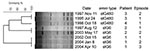
Figure 2. Dendrogram and pulsed-field gel electrophoresis (PFGE) profiles of the strains isolated from patients with recurrent group G Streptococcus dysgalagtiae subsp. equisimilis bacteremia, Finland. Dendogram was generated by using Bionumerics software (Applied...
We divided bacteremia episodes into 2 groups: those caused by the 5 most common emm types and each representing >5% of all episodes (97 episodes, common emm types) and those caused by the less common or nontypable emm types (41 episodes, rare emm types). We could not find an association between emm type and clinical features such as age or underlying disease. Severe disease was caused more often by rare emm types than by common emm types. Mortality rates were higher in patients with bacteremia caused by rare types than that caused by common types (Table 1). Four patients had recurrent S. dysgalactiae subsp. equisimilis bacteremia (Table 2). PFGE profiles showed that strains isolated from the same patient in recurring infections were identical (Figure 2).
Common emm types were more frequently manifested as skin and soft tissue infections than were rare emm types, 75% vs. 54%, respectively (p = 0.012). The most frequent source of bacteremia was cellulitis (51%). We also found an association between a common emm type and cellulitis. Cellulitis was a more frequent clinical sign among patients with infections caused by common emm types than by rare emm types (p = 0.007); 64% of patients infected by common emm types had cellulitis as an initial clinical manifestation versus 39% of patients infected by rare emm types.
Our study showed that mortality rates were higher in patients with S. dysgalactiae subsp. equisimilis bacteremia caused by rare emm types than in those with bacteremia caused by common emm types. The reason for this finding is unclear. One explanation for this might be that patients contract certain prevailing bacterial strains (so-called common types) more often, and a prior antigen challenge and subsequent humoral response may play a role. Severe disease (death or intensive care unit treatment) was also caused more often by rare emm types than by common emm types. We found also an association between a common emm type and cellulitis as a clinical manifestation; the common emm types were also associated with skin and soft tissue infections.
In our comprehensive study with molecular typing data for 138 invasive S. dysgalactiae subsp. equisimilis isolates from human infections, we found 18 emm types, which is consistent with previous reports by Cohen-Poradosu et al. (12) and Broyles et al. (13) . These 2 studies reported stG485.0. or StG6, StG245, and StG2078 as the most common emm types, respectively. Thus, emm typing provides a useful tool for comparative epidemiologic analysis of GGS isolates from various geographic regions. Our results also suggest that certain emm types may prevail among bacteria that cause human infections. Our study did not show any obvious time shifts in the occurrence of certain emm types.
A noteworthy finding in our series was the high frequency of recurrent group G S. dysgalactiae subsp. equisimilis bacteremia as reported earlier (12,14). Clinicians should be alert to this phenomenon, which seems to be more common than recurrent group A streptococcal bacteremia.
The dynamics of interspecies transfer of virulence loci between group A streptococci, GGS, and GCS (3), as well as potential genetic transfer or intragenomic events causing interconversion of group antigen types, remains to be resolved. Further characterization of the strains by multilocus sequence typing would be of interest (15).
We conclude that severity of disease and mortality rates were higher in persons with S. dysgalactiae subsp. equisimilis bacteremia caused by rare emm types than that caused by common emm types. Skin and soft tissue infections such as cellulitis were significantly more frequent clinical signs among episodes caused by common emm types.
Dr Rantala is a specialist in infectious disease and internal medicine in the Department of Internal Medicine, Tampere University Hospital, and a consultant in infectious diseases at Tampere University Hospital. Her research interests include streptococcal infections.
Acknowledgments
We thank Esko Väyrynen for the revision of the language in this article.
This work was supported by a grant from the Medical Research Fund of Tampere University Hospital. The study was presented in part at the 18th European Congress of Clinical Microbiology and Infectious Diseases, Barcelona, Spain, April 2008 (poster no. P 1824).
References
- Vartian C, Lerner PI, Shlaes DM, Gopalakrishna KV. Infections due to Lancefield group G streptococci. Medicine (Baltimore). 1985;64:75–88. DOIPubMedGoogle Scholar
- Facklam R. What happened to the streptococci: overview of taxonomic and nomenclature changes. Clin Microbiol Rev. 2002;15:613–30. DOIPubMedGoogle Scholar
- Brandt CM, Spellerberg B. Human infections due to Streptococcus dysgalactiae subspecies equisimilis. Clin Infect Dis. 2009;49:766–72. DOIPubMedGoogle Scholar
- Bisno AL, Craven DE, McCabe WR. M proteins of group G streptococci isolated from bacteremic human infections. Infect Immun. 1987;55:753–7.PubMedGoogle Scholar
- Lancefield RC. Current knowledge of type-specific M antigens of group A streptococci. J Immunol. 1962;89:307–13.PubMedGoogle Scholar
- Facklam R, Beall B, Efstratiou A, Fischetti V, Johnson D, Kaplan E, emm typing and validation of provisional M types for group A streptococci. Emerg Infect Dis. 1999;5:247–53. DOIPubMedGoogle Scholar
- Bisno AL, Collins CM, Turner JC. M proteins of group C streptococci isolated from patients with acute pharyngitis. J Clin Microbiol. 1996;34:2511–5.PubMedGoogle Scholar
- Rantala S, Vuopio-Varkila J, Vuento R, Huhtala H, Syrjanen J. Clinical presentations and epidemiology of beta-haemolytic streptococcal bacteraemia: a population-based study. Clin Microbiol Infect. 2009;15:286–8. DOIPubMedGoogle Scholar
- Siljander T, Toropainen M, Muotiala A, Hoe NP, Musser JM, Vuopio-Varkila J. emm typing of invasive T28 group A streptococci, 1995–2004, Finland. J Med Microbiol. 2006;55:1701–6. DOIPubMedGoogle Scholar
- Siljander T, Karppelin M, Vahakuopus S, Syrjanen J, Toropainen M, Kere J, Acute bacterial, nonnecrotizing cellulitis in Finland: microbiological findings. Clin Infect Dis. 2008;46:855–61. DOIPubMedGoogle Scholar
- Tenover FC, Arbeit RD, Goering RV, Mickelsen PA, Murray BE, Persing DH, Interpreting chromosomal DNA restriction patterns produced by pulsed-field gel electrophoresis: criteria for bacterial strain typing. J Clin Microbiol. 1995;33:2233–9.PubMedGoogle Scholar
- Cohen-Poradosu R, Jaffe J, Lavi D, Grisariu-Greenzaid S, Nir-Paz R, Valinsky L, Group G streptococcal bacteremia in Jerusalem. Emerg Infect Dis. 2004;10:1455–60.PubMedGoogle Scholar
- Broyles LN, Van Beneden C, Beall B, Facklam R, Shewmaker PL, Malpiedi P, Population-based study of invasive disease due to beta-hemolytic streptococci of groups other than A and B. Clin Infect Dis. 2009;48:706–12. DOIPubMedGoogle Scholar
- Liao CH, Liu LC, Huang YT, Teng LJ, Hsueh PR. Bacteremia caused by group G streptococci, Taiwan. Emerg Infect Dis. 2008;14:837–40. DOIPubMedGoogle Scholar
- Ahmad Y, Gertz RE Jr, Li Z, Sakota V, Broyles LN, Van Beneden C, Genetic relationships deduced from emm and multilocus sequence typing of invasive Streptococcus dysgalactiae subsp. equisimilis and S. canis recovered from isolates collected in the United States. J Clin Microbiol. 2009;47:2046–54. DOIPubMedGoogle Scholar
Figures
Tables
Cite This ArticleTable of Contents – Volume 16, Number 5—May 2010
| EID Search Options |
|---|
|
|
|
|
|
|
Please use the form below to submit correspondence to the authors or contact them at the following address:
Sari Rantala, Department of Internal Medicine, Tampere University Hospital, PO Box 2000, FIN–33521, Tampere, Finland
Top