Volume 17, Number 3—March 2011
Letter
Risk for Mycobacterium celatum Infection from Ferret
Figure
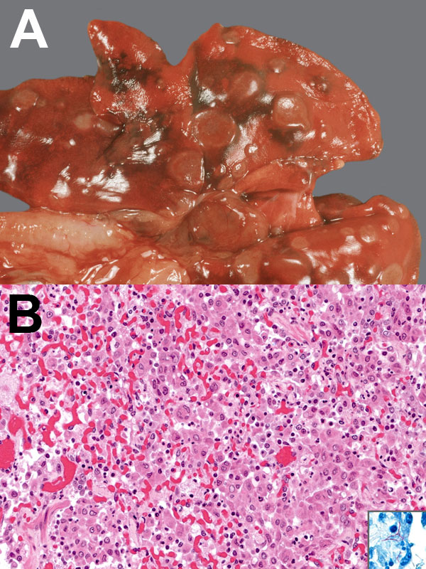
Figure. Appearance of tissue from 3-year-old, neutered male, domestic ferret with Mycobacterium celatum infection. A) Gross appearance: multiple, round light brown foci over lungs. B) Histologic appearance, granulomatous pneumonia: alveoli filled with foamy macrophages, epithelioid cells, and a multinucleated giant cell; also mild interstitial infiltration with lymphocytes, plasma cells, and neutrophils. Hematoxylin and eosin staining, original magnification x200. Inset, slender, rod-shaped, acid-fast bacilli in the cytoplasm of epithelioid cells; Ziehl-Neelsen staining, original magnification x400. A color version of this figure is available online (www.cdc.gov/EID/content/17/3/553-F.htm).
1These authors contributed equally to this article.