Volume 19, Number 5—May 2013
Dispatch
Detecting Rickettsia parkeri Infection from Eschar Swab Specimens
Todd Myers , Tahaniyat Lalani, Mike Dent, Ju Jiang, Patrick L. Daly, Jason D. Maguire, and Allen L. Richards
Figure
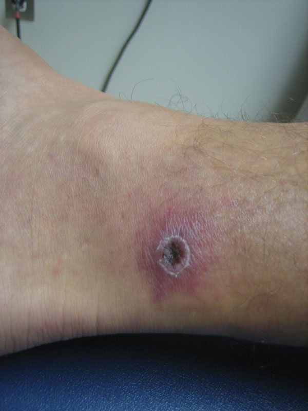
Figure. . . Acute eschar of patient who was subsequently diagnosed with Rickettsia parkeri infection in Pensacola, Florida, USA, in August 2011. This same eschar was unsheathed and swabbed after 14 days of antimicrobial drug treatment and had undergone significant healing. It still gave a positive result by real time PCR, although the convalescent-phase blood specimen showed a negative result.
Page created: April 03, 2013
Page updated: April 03, 2013
Page reviewed: April 03, 2013
The conclusions, findings, and opinions expressed by authors contributing to this journal do not necessarily reflect the official position of the U.S. Department of Health and Human Services, the Public Health Service, the Centers for Disease Control and Prevention, or the authors' affiliated institutions. Use of trade names is for identification only and does not imply endorsement by any of the groups named above.