Volume 20, Number 2—February 2014
Dispatch
Molecular Detection of Diphyllobothrium nihonkaiense in Humans, China
Shaohong Chen1, Lin Ai, Yongnian Zhang, Jiaxu Chen, Weizhe Zhang1, Yihong Li, Maki Muto1, Yasuyuki Morishima, Hiromu Sugiyama, Xuenian Xu1, Xiaonong Zhou1, and Hiroshi Yamasaki1
Figure 1
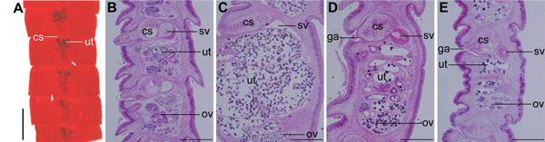
Figure 1. Diphyllobothriid samples examined in the present study, China, 2008–2012A) Proglottids stained with acetic acid–carmine from case-patient 12B–E) Sagittal sections of proglottids stained with hematoxylin-eosin from case-patients 16–19cs, cirrus sac; ut, uterus; sv, seminal vesicle; ov, ovary; ga, genital atriumScale bar in panel A represents 2 mm; scale bars in panels B–E represent 500 μm.
1These authors contributed equally to this article.
Page created: January 16, 2014
Page updated: January 16, 2014
Page reviewed: January 16, 2014
The conclusions, findings, and opinions expressed by authors contributing to this journal do not necessarily reflect the official position of the U.S. Department of Health and Human Services, the Public Health Service, the Centers for Disease Control and Prevention, or the authors' affiliated institutions. Use of trade names is for identification only and does not imply endorsement by any of the groups named above.