Volume 20, Number 4—April 2014
Research
Antibodies against MERS Coronavirus in Dromedary Camels, United Arab Emirates, 2003 and 2013
Benjamin Meyer, Marcel A. Müller, Victor M. Corman, Chantal B.E.M. Reusken, Daniel Ritz, Gert-Jan Godeke, Erik Lattwein, Stephan Kallies, Artem Siemens, Janko van Beek, Jan F. Drexler, Doreen Muth, Berend-Jan Bosch, Ulrich Wernery, Marion P.G. Koopmans, Renate Wernery, and Christian Drosten
Figure
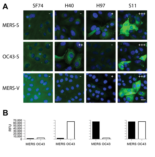
Figure. . . Immunofluorescence and microarray reactivity patterns for antibodies (SF74, H40, H97, and S11) against Middle East respiratory syndrome coronavirus (MERS-CoV) in serum samples from camels, United Arab Emirates, 2013. A) Serum samples tested against overexpressed MERS-CoV spike protein (MERS-S), overexpressed human CoV-OC43 spike protein (OC43-S), and Vero cells infected with MERS-CoV (MERS-V). Fluorescence intensities were evaluated as follows: –, negative; +, weakly reactive; ++, reactive; +++, strongly reactive. Scale bar indicates 20 μm. B) Relative fluorescence units (RFU) were determined for the same serum samples by microarray using S1 domains of MERS-CoV and human CoV-OC43.
Page created: March 12, 2014
Page updated: March 12, 2014
Page reviewed: March 12, 2014
The conclusions, findings, and opinions expressed by authors contributing to this journal do not necessarily reflect the official position of the U.S. Department of Health and Human Services, the Public Health Service, the Centers for Disease Control and Prevention, or the authors' affiliated institutions. Use of trade names is for identification only and does not imply endorsement by any of the groups named above.