Volume 27, Number 11—November 2021
Research Letter
Co-Infection with 4 Species of Mycobacteria Identified by Using Next-Generation Sequencing
Abstract
We identified co-infection with 4 species of mycobacteria in a woman in New York, New York, USA, by using next-generation sequencing. This procedure is useful for identifying co-infections with multiple mycobacteria, tracing the geographic origin of strains, investigating transmission dynamics in susceptible populations, and gaining insight into prevention and control.
Mycobacteria are major human pathogens; ≈13 million persons in the United States live with Mycobacterium tuberculosis complex (MTBC) infection, and incidence of nontuberculous mycobacterial (NTM) pulmonary disease is increasing worldwide. The challenges of managing MTBC and M. avium complex (MAC) co-infection are well described, including the risk for falsely interpreted Xpert RIF (rifampin) results (1,2). MTBC and M. abscessus co-infection has been described in case reports only (3,4). We describe co-infection with 4 species of mycobacteria.
In July 2019, an 82-year-old Asian woman was hospitalized in Flushing, New York, USA, for persistent fever associated with worsening weakness. Computed tomography of her chest showed near-complete atelectasis of the left upper lobe, hyperinflation in other areas, and a small left-sided pleural effusion. Scattered nodular opacities in a tree-in-bud pattern and pulmonary granulomas were present throughout the lungs, and discontinuity of the left upper lobe bronchus was noted. Cultures of blood, urine, stool, and respiratory specimens yielded negative results for nonmycobacteria.
In a sputum sample collected for routine mycobacterial testing, fluorochrome staining exhibited rare acid-fast bacilli, and MTBC was detected by using Xpert MTB/RIF (Cepheid, https://www.cepheid.com). We then inoculated a Lowenstein-Jensen Gruft slant with sputum, incubated it at 37°C, and inoculated VersaTREK Myco bottles containing Modified Middlebrook 7H9 Broth with Sponges (Thermo Fisher, https://www.thermofisher.com) and incubated them at 35°C. No isolate was recovered from the Lowenstein-Jensen Gruft slant. Only MAC was detected by AccuProbe (Hologic, https://www.hologic.com) in Kinyoun-positive culture from the Myco bottles. One week later, another sputum sample with Kinyoun-positive growth from the Myco bottles was negative for MAC, MTBC, M. gordonae, and M. kansasii. M. abscessus was identified on the Lowenstein-Jensen Gruft slant.
Considering the sensitivity limit and narrow species coverage of AccuProbe and the difficulty of identifying mycobacteria by culturing and because of growth interference among different mycobacteria, we conducted next-generation sequencing (NGS) by using Hiseq3000 (Illumina, https://www.illumina.com) on the supernatant of the first sputum culture. NGS yielded ≈175 million reads, each with a quality score of >35. We checked NGS data for quality control by using FastQC (Galaxy, https://usegalaxy.org). All steps and programs used the data processing pipeline from Galaxy, an open-source, web-based platform for data-intensive biomedical research. Each read identified had a quality control score of 39.4 and an average guanine-cytosine content of 68%. Only 0.69% of bases resulted in no hits and were not identifiable. We performed De Novo classification by using De Novo Assembly Unicycler, Quast QC, and Kraken Classification (Galaxy) and generated coverage and depth data by using BWA Aligner and SAMtools Depth (Galaxy). We aligned the reads, visualized onto bacteria reference genomes by using Bowtie2 (Galaxy) and converted into BED (Browser Extensible Data) files followed by Bedtools Merge, Multicov (https://bedtools.readthedocs.io).
Figure
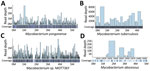
Figure. Genomic coverage and depth map of 4 Mycobacteriumstrains identified by using next-generation sequencing on isolates from a woman in New York, New York, USA. The reads were aligned...
The genome visualization pipeline confirmed 4 genomic traces of Mycobacterium strains (Figure): M. yongonense strain 05-1390 (GenBank accession no. NC_021715.1), M. tuberculosis strain FDAARGOS_757 (GenBank accession no. CP054013.1), Mycobacterium sp. MOTT36Y (GenBank accession no. NC_017904.1), and M. abscessus ATCC 19977 (GenBank accession no. CU458896.1). M. yongonense was identified with a genome coverage of 88.73% (4.9 Mb mapped of 5.5-Mb genome) and a read depth of 1,224×. M. tuberculosis was identified with a genome coverage of 99.99% (4.4 Mb mapped of 4.4-Mb genome) and a read depth of 63×. Mycobacterium sp. was identified with a genome coverage of 94.41% (5.3 Mb mapped of 5.6-Mb genome) and a read depth of 1210×. M. abscessus was identified with a genome coverage of only 2.75% (0.14 Mb mapped of 5.1-Mb genome) and a read depth of 8× (Table). The mycobacteria identified by NGS were verified by various mycobacteria tests.
We obtained the consensus sequence for 4 strains of bacteria by using MEGAHIT (Galaxy) and generated a BLAST (https://blast.ncbi.nlm.nih.gov/Blast.cgi) tree based on minimum evolution at the species level by using >15 kbp from each sequence. Assembly on MTBC sequencing data yielded a total consensus sequence of 4,376,826 bp and 78,208 single-nucleotide polymorphism sites (1.79%). Analysis by BLAST and Mykrobe (https://www.mykrobe.com) revealed that the MTBC isolate belongs to sublineage 2.2.
The patient received RIPE therapy (rifampin, isoniazid, pyrazinamide, and ethambutol), along with amikacin, tigecycline, and azithromycin. At 6 months, RIPE therapy was completed. At 9 months, sputum culture was negative. The patient continues to take amikacin, tigecycline, and azithromycin as an outpatient with close follow-up.
Identification of co-infection with mycobacteria is necessary for diagnosis and treatment (5). Treatment regimens and duration remain species specific because of unique resistance mechanisms. To achieve the greatest potential for success while minimizing toxicities, early empiric treatment should account for clinical characteristics of MTBC and NTM co-infection and strain identification.
Our report highlights the value of NGS for identifying multiple mycobacteria co-infections in populations with high susceptibility to and prevalence of MTB and NTM (i.e., immigrants, immunocompromised patients, and international travelers). NGS can trace the geographic origin of the Mycobacterium strain. These features, in combination with a patient’s epidemiologic exposure and travel history, could elucidate the potential time and location of infection acquisition. NGS could also be used to identify drug-resistance genes to guide targeted therapy.
Dr. Wang is a postdoctoral fellow working in the Department of Microbiology, Immunology and Molecular Genetics, University of California, Los Angeles. His research focuses on immunology, virology, and molecular diagnostics. Dr. Liu is the laboratory director for Microbiology, Immunology and Molecular Diagnostics, Department of Pathology and Clinical Laboratories, New York-Presbyterian Queens Hospital. His research focus is developing rapid molecular assays for pathogen diagnosis.
Acknowledgments
We thank Jun Young Choi, Debra Harragan Jokisch, Eric Konadu, Marian Memmer, Joseph Pilz, Huimin Wu for their technical assistance and Carl Urban for manuscript review.
The sequenced isolate in this manuscript was submitted to GenBank (accession no. CP074075). Details of submission are SUBID SUB9558401; BioProject PRJNA726345: BioSample SAMN18928235; Organism Mycobacterium tuberculosis 2.2.
References
- Bazzi AM, Abulhamayel Y, Rabaan AA, Al-Tawfiq JA. The impact of the coexistence of mycobacterium avium with mycobacterium tuberculosis on the result of GeneXpert and MGIT susceptibility test. J Infect Public Health. 2020;13:827–9. DOIPubMedGoogle Scholar
- Sarro YDS, Kone B, Diarra B, Kumar A, Kodio O, Fofana DB, et al. Simultaneous diagnosis of tuberculous and non-tuberculous mycobacterial diseases: Time for a better patient management. Clin Microbiol Infect Dis. 2018;3:1–8. DOIPubMedGoogle Scholar
- Sohn S, Wang S, Shi H, Park S, Lee S, Park KT. Mixed Infection of Mycobacterium abscessus subsp. abscessus and Mycobacterium tuberculosis in the Lung. Korean J Thorac Cardiovasc Surg. 2017;50:50–3. DOIPubMedGoogle Scholar
- Ishiekwene C, Subran M, Ghitan M, Kuhn-Basti M, Chapnick E, Lin YS. Case report on pulmonary disease due to coinfection of Mycobacterium tuberculosis and Mycobacterium abscessus: Difficulty in diagnosis. Respir Med Case Rep. 2017;20:123–4. DOIPubMedGoogle Scholar
- Feng Z, Bai X, Wang T, Garcia C, Bai A, Li L, et al. Differential responses by human macrophages to infection with Mycobacterium tuberculosis and non-tuberculous mycobacteria. Front Microbiol. 2020;11:116. DOIPubMedGoogle Scholar
Figure
Table
Cite This ArticleOriginal Publication Date: October 08, 2021
1These first authors contributed equally to this article.
Table of Contents – Volume 27, Number 11—November 2021
| EID Search Options |
|---|
|
|
|
|
|
|
Please use the form below to submit correspondence to the authors or contact them at the following address:
Dakai Liu and William H. Rodgers, Department of Pathology and Clinical Laboratories, New York Presbyterian Queens Hospital, 56-45 Main St., Flushing, NY 11355, USA
Top