Volume 31, Number 11—November 2025
Research Letter
Crimean-Congo Hemorrhagic Fever Virus in Cattle and Ticks, Israel
Cite This Article
Citation for Media
Abstract
We conducted a nationwide serologic and molecular survey to elucidate the epidemiologic status of Crimean-Congo hemorrhagic fever virus in Israel. We found serologic and molecular evidence of virus circulation in the country. Future human cases could be prevented by increasing public awareness and implementing public health measures.
Crimean-Congo hemorrhagic fever virus (CCHFV) is an enveloped segmented negative-sense RNA virus belonging to the Nairoviridae family of the Bunyavirales order (1). The virus is the etiologic agent of Crimean-Congo hemorrhagic fever (CCHF), a severe tickborne zoonotic illness with a wide geographic distribution, infecting ≈10,000–15,000 humans annually worldwide (2). Ticks, primarily of the genus Hyalomma, are considered the vector and the reservoir of CCHFV (3). CCHFV can infect various animal hosts, including livestock, that, although remaining asymptomatic, can act as amplifying hosts of the virus (4). CCHFV is transmitted to humans primarily through the bite of an infected tick but also by direct contact with blood or body fluids of infected animals or humans or through improperly sterilized medical equipment (1).
Neither CCHFV infection in humans nor positive serologic test results in humans or in animals were previously reported in Israel. However, outbreaks of the disease and seropositivity among livestock have been reported in neighboring countries (5). Moreover, the main vector of CCHFV, the Hyalomma marginatum tick, is endemic in Israel (6). Hence, the risk for CCHFV emergence in Israel is considered high, and undetected viral circulation might already exist in specific regions of the country (6).
During April 2024–February 2025, we sampled whole blood, serum specimens, and ticks from 19 beef cattle herds from different regions in Israel. We tested serum samples by using an ELISA commercial kit (ID Screen CCHF Double Antigen Multi-species; Innovative Diagnostics, https://www.innovative-diagnostics.com). We classified the ticks morphologically and extracted RNA to identify CCHFV presence (Appendix).
Figure
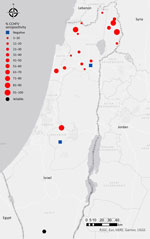
Figure. Distribution of samples seropositive for CCHFV in study of serologic and molecular evidence of CCHFV in cattle and ticks, Israel, April 2024–February 2025. Red dots represent seropositive beef cattle herds....
Sixteen beef cattle herds had serologic evidence of prior exposure to CCHFV; seropositivity ranged from 3% to 100% (Table). We detected virus exposure eliciting an immune response in locations across Israel (Figure). Those serologic results are comparable with rates reported in CCHFV-endemic countries with confirmed human cases, such as Turkey (Türkiye) (7) and Pakistan (8). Consistent with prior publications linking older age with seropositivity (9), the age of the sampled animals from the 2 herds that were seronegative was <2 years. Moreover, although all serum samples from the heifers (<2 years of age) of the Keshet herd (Golan Heights) were seronegative, subsequent sampling of older cows (3–15 years of age) in the herd revealed 100% positive serologic test results (Table). We also tested serum samples that were randomly collected from 200 wild animals dispersed through the country (including boars, foxes, jackals, ibexes, dogs, deer, oryxes, gazelles, and porcupines) during 2023–2024. We found that only 2 samples, from ibexes (Capra nubiana) residing in the Negev Desert (southern Israel), were seropositive for CCHFV antibodies (Figure).
We tested for CCHFV RNA in 227 ticks retrieved from 8 cattle herds and 51 ticks retrieved from wildlife by using quantitative reverse transcription PCR targeting 2 regions of the small segment (10). Because we tested each tick individually, we determined a sample to be positive only when both regions were amplified. In addition, the positive samples were validated at an independent facility (The Central Virology Laboratory, Ministry of Health and Sheba Medical Center, Ramat-Gan, Israel). Of the 227 ticks from cattle, 23 (10%) samples collected from northern Israel (Golan Heights and Western Galilee) were positive for CCHFV (Appendix Table 1). Likewise, among 51 ticks collected from wild animals (all sampled from northern Israel), 7 (13%) were positive for CCHFV (Appendix Table 2). All ticks positive for CCHFV belonged to 2 genera, Hyalomma and Rhipicephalus (Appendix Tables 1, 2). Sanger sequencing of the 181-bp (1,068–1,248 nucleotides at the small segment) amplicons of 10 samples, which we successfully amplified by using endpoint reverse transcription PCR (10), followed by phylogenetic analysis, indicated that the sequences clustered to the Asia-1 genotype (Appendix Figure).
We tested serum samples from 13 persons who had close contact with either CCHFV-positive ticks or seropositive cattle for the presence of CCHF IgG by using ELISA (Euroimmun, https://www.euroimmun.com). All persons tested were seronegative. The study was conducted with the approval of the Shiba Medical Center institutional review board (approval no. 1601–24-SMC).
We present evidence of CCHFV circulation in Israel, expanding the known geographic distribution of CCHFV in the Middle East. We have found serologic evidence of prior exposure to CCHFV in livestock and in wild animals; we also detected CCHFV in the ticks infesting them. Furthermore, the seroprevalence in the cows was found to be comparable to seroprevalence in CCHFV-endemic countries with proven human cases. In addition, the seropositivity prevalence in the cattle and the observation that cows <2 years of age were not seropositive suggest that the virus has been circulating for several years. Still, the route of introduction remains unclear. Our results highlight the importance of raising public and clinical awareness of CCHF, especially among high-risk populations, despite the current absence of human cases.
Dr. Rudoler is head of the virology laboratory at the Kimron Veterinary Institute, Israel. His major interests include veterinary public health, zoonotic diseases, and conducting and implementing One Health research projects.
Acknowledgment
We thank the persons who helped to collect or analyze serum samples or ticks for this project, including Adi Behar, Monica Leszkowicz-Mazuz, Meytal Bakal-Weiss, Geva Amaton, Boris Even-Tov, Lior Zamir, Hamed Fares, Yuval Hadani, Ricardo Wolkomirsky, Sharon Karniely, Nick Storm, Roni King, Roi Lapid, Tomer Nissimyan, and Schahar Ertracht. We also extend our gratitude to the cattle farmers who patiently collaborated with us and gave us access to their facilities and cattle.
References
- Hawman DW, Feldmann H. Crimean-Congo haemorrhagic fever virus. Nat Rev Microbiol. 2023;21:463–77. DOIPubMedGoogle Scholar
- Frank MG, Weaver G, Raabe V; State of the Clinical Science Working Group of the National Emerging Pathogens Training and Education Center’s Special Pathogens Research Network. Crimean-Congo hemorrhagic fever virus for clinicians—virology, pathogenesis, and pathology. Emerg Infect Dis. 2024;30:847–53. DOIPubMedGoogle Scholar
- Gargili A, Estrada-Peña A, Spengler JR, Lukashev A, Nuttall PA, Bente DA. The role of ticks in the maintenance and transmission of Crimean-Congo hemorrhagic fever virus: a review of published field and laboratory studies. Antiviral Res. 2017;144:93–119. DOIPubMedGoogle Scholar
- Spengler JR, Bergeron É, Rollin PE. Seroepidemiological studies of Crimean-Congo hemorrhagic fever virus in domestic and wild animals. PLoS Negl Trop Dis. 2016;10:
e0004210 . DOIPubMedGoogle Scholar - Perveen N, Khan G. Crimean-Congo hemorrhagic fever in the Arab world: a systematic review. Front Vet Sci. 2022;9:
938601 . DOIPubMedGoogle Scholar - Messina JP, Wint GRW. The spatial distribution of Crimean-Congo haemorrhagic fever and its potential vectors in Europe and beyond. Insects. 2023;14:771. DOIPubMedGoogle Scholar
- Nurettin C, Engin B, Sukru T, Munir A, Zati V, Aykut O. The seroprevalence of Crimean-Congo hemorrhagic fever in wild and domestic animals: an epidemiological update for domestic animals and first seroevidence in wild animals from Turkiye. Vet Sci. 2022;9:462. DOIPubMedGoogle Scholar
- Zohaib A, Saqib M, Athar MA, Hussain MH, Sial AU, Tayyab MH, et al. Crimean-Congo hemorrhagic fever virus in humans and livestock, Pakistan, 2015–2017. Emerg Infect Dis. 2020;26:773–7. DOIPubMedGoogle Scholar
- Schulz A, Barry Y, Stoek F, Ba A, Schulz J, Haki ML, et al. Crimean-Congo hemorrhagic fever virus antibody prevalence in Mauritanian livestock (cattle, goats, sheep and camels) is stratified by the animal’s age. PLoS Negl Trop Dis. 2021;15:
e0009228 . DOIPubMedGoogle Scholar - Mazzola LT, Kelly-Cirino C. Diagnostic tests for Crimean-Congo haemorrhagic fever: a widespread tickborne disease. BMJ Glob Health. 2019;4(Suppl 2):
e001114 . DOIPubMedGoogle Scholar
Figure
Table
Cite This ArticleOriginal Publication Date: November 24, 2025
Table of Contents – Volume 31, Number 11—November 2025
| EID Search Options |
|---|
|
|
|
|
|
|
Please use the form below to submit correspondence to the authors or contact them at the following address:
Elad Eliahoo, Virology Diagnostic Laboratory, Department of Virology, Kimron Veterinary Institute, POB 30, Beit Dagan 5025001, Israel
Top