Volume 13, Number 11—November 2007
Research
Non-A Hepatitis B Virus Genotypes in Antenatal Clinics, United Kingdom
Samir Dervisevic* , Samreen Ijaz†, Shahneila Chaudry*, and Richard S. Tedder*†
Figure
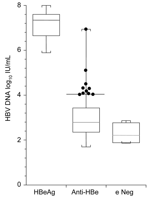
Figure. Box and whisker plots of hepatitis B virus (HBV) load in 3 groups of mothers whose serum contained hepatitis B virus e antigen (HBeAg), antibody to hepatitis B virus e antigen (anti-HBe), or neither of these markers (e Neg). Boxes are middle quartiles, horizontal lines are medians, whiskers are ranges, and dots represent 10 anti-HBe–seropositive mothers whose serum contained >104 IU/mL HBV DNA. Thirty-three anti-HBe–seropositive mothers and 1 mother whose serum did not contain either marker did not have detectable HBV DNA (<50 IU/mL).
Page created: July 06, 2010
Page updated: July 06, 2010
Page reviewed: July 06, 2010
The conclusions, findings, and opinions expressed by authors contributing to this journal do not necessarily reflect the official position of the U.S. Department of Health and Human Services, the Public Health Service, the Centers for Disease Control and Prevention, or the authors' affiliated institutions. Use of trade names is for identification only and does not imply endorsement by any of the groups named above.