Volume 14, Number 12—December 2008
Letter
Human Case of Bartonella alsatica Lymphadenitis
Appendix Figure
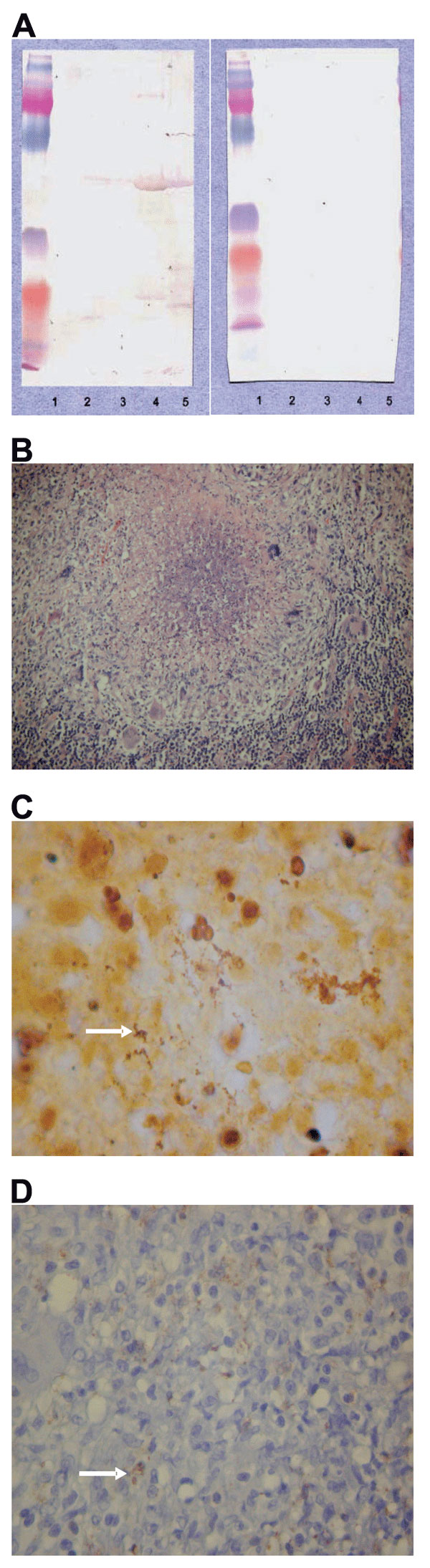
Appendix Figure. A) Western blotting analysis of lymph node specimen from the patient before 1) and after 2) cross-adsorption with Bartonella alsatica. Lane 1, B. quintana; lane 2, B. henselae; lane 3, B. elizabethae; lane 4, B. vinsonii subsp. berkhoffii; lane 5, B. alsatica. B) Characteristic histologic change in the lymph node with B. alsatica infection. Shown is an inflammatory granulomatous process with central microabscess surrounded by a ring of macrophages and rare giant cells (hematoxylin and eosin stain, original magnification x100). C) Bacteria (arrow) in an abscess formation mixed with necrotic debris (Warthin-Starry silver stain, original magnification x1,000). D) Immunohistochemical detection of B. alsatica (arrow) in lymph node pulp with an extracellular distribution (polyclonal antibody and hematoxylin counterstain, original magnification x400).