Volume 15, Number 4—April 2009
Dispatch
Co-infection with Pansensitive and Multidrug-Resistant Strains of Mycobacterium tuberculosis
Michael P. Mendez , Mary E. Landon, Mary K. McCloud, Peter Davidson, and Paul J. Christensen
Figure
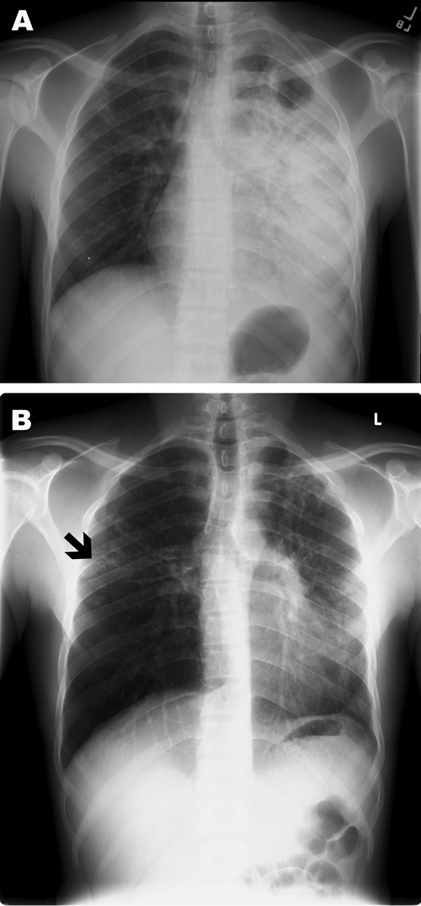
Figure. Poster-anterior chest radiographs of patient with multidrug-resistant tuberculosis. A) Radiograph taken at diagnosis, demonstrating dense consolidation of the left lower lobe and lingula. A left apical cavity is present. Minimal change is also noted in the right mid-lung zone. B) Radiograph taken after 5 months of directly observed therapy. Marked clearance is noted on the left; however, a new small cavitary lesion with surrounding infiltrate is noted in the right mid-lung zone (black arrow).
Page created: December 10, 2010
Page updated: December 10, 2010
Page reviewed: December 10, 2010
The conclusions, findings, and opinions expressed by authors contributing to this journal do not necessarily reflect the official position of the U.S. Department of Health and Human Services, the Public Health Service, the Centers for Disease Control and Prevention, or the authors' affiliated institutions. Use of trade names is for identification only and does not imply endorsement by any of the groups named above.