Volume 5, Number 3—June 1999
Synopsis
Emergence of a Unique Group of Necrotizing Mycobacterial Diseases
Karen M. Dobos*†, Frederick D. Quinn†, David A. Ashford†, C. Robert Horsburgh*, and C. Harold King*
Figure 3 A and B
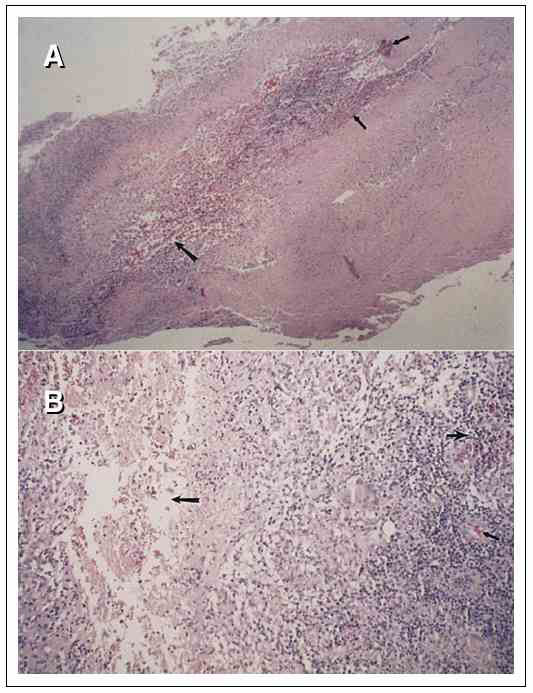
Figure 3 A and B. Active disease histopathologic sections of the epidermis stained for acid-fast bacilli from a patient infected with Mycobacterium haemophilum. In A and B, the arrows indicate localized necrosis and presence of intracellular and extracellular bacilli and microcolonies and the presence of loose granulomas. (Slide courtesy of Michael A. Saubolle, Good Samaritan Regional Medical Center, Phoenix, Arizona, and Department of Medicine, University of Arizona School of Medicine, Tucson, Arizona.)
Page created: December 10, 2010
Page updated: December 10, 2010
Page reviewed: December 10, 2010
The conclusions, findings, and opinions expressed by authors contributing to this journal do not necessarily reflect the official position of the U.S. Department of Health and Human Services, the Public Health Service, the Centers for Disease Control and Prevention, or the authors' affiliated institutions. Use of trade names is for identification only and does not imply endorsement by any of the groups named above.