Volume 9, Number 6—June 2003
Research
Histopathologic Features of Mycobacterium ulcerans Infection
Jeannette Guarner* , Jeanine Bartlett*, Ellen A. Spotts Whitney*, Pratima L. Raghunathan*, Ymkje Stienstra†, Kwame Asamoa‡, Samuel Etuaful§, Erasmus Klutse¶, Eric Quarshie#, Tjip S. van der Werf†, Winette T.A. van der Graaf†, C. Harold King**, and David A. Ashford*
Figure 2
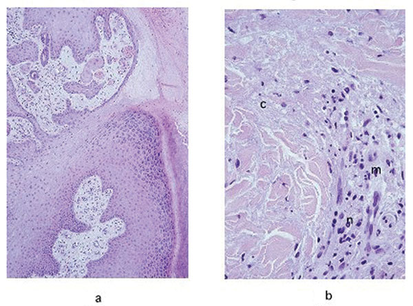
Figure 2. a, hematoxylin and eosin stain of the pseudoepitheliomatous hyperplasia of the epidermis in a lesion specimen showing definitive Buruli ulcer disease in the ulcerative stage (original magnification 100x). b, hematoxylin and eosin stain of the necrotic collagen (c) accompanied by mild inflammatory infiltrate in the dermis of a definitive Buruli ulcer disease lesion in the ulcerative stage (original magnification 400x). n, neutrophis; m, mononuclear cells.
Page created: December 21, 2010
Page updated: December 21, 2010
Page reviewed: December 21, 2010
The conclusions, findings, and opinions expressed by authors contributing to this journal do not necessarily reflect the official position of the U.S. Department of Health and Human Services, the Public Health Service, the Centers for Disease Control and Prevention, or the authors' affiliated institutions. Use of trade names is for identification only and does not imply endorsement by any of the groups named above.