Volume 23, Number 7—July 2017
Research Letter
Mycobacterium gordonae in Patient with Facial Ulcers, Nosebleeds, and Positive T-SPOT.TB Test, China
Figure
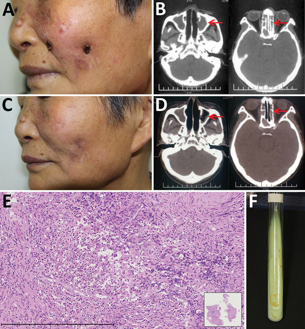
Figure. Mycobacterium gordonae infection in a 60-year-old immunocompetent woman, China. A) Facial lesions before treatment. Ulcers were erythematous and covered with yellow crusts. B) Computed tomography images before treatment show heterogeneous hypersignal in the ethmoid and left maxillary sinus (arrows). C) Facial lesions after treatment. Atrophic scars are seen at sites of previous lesions. D) Computed tomography images after treatment show recovery of the ethmoid sinus and left maxillary sinus (arrows). E) Hematoxylin and eosin stain of nasal mucosa showing the infiltration of a large number of lymphocytes, a few histiocytes, and plasma cells. Scale bar corresponds to 400 µm. Inset shows the nasal mucosa sample (original magnification ×20). F) Tissue culture 3 weeks after incubation shows yolk-yellow bacteria growing in Löwenstein–Jensen medium. A color version of this figure is available online (https://wwwnc.cdc.gov/EID/article/23/7/16-2033-F1.htm).