Skip directly to site content Skip directly to page options Skip directly to A-Z link Skip directly to A-Z link Skip directly to A-Z link
Volume 23, Number 7—July 2017
Research Letter

Mycobacterium gordonae in Patient with Facial Ulcers, Nosebleeds, and Positive T-SPOT.TB Test, China

Yanqing Chen, Juan Jiang, Haiqin Jiang, Jun Chen, Xiaopo Wang, Weida Liu, Zhiming Chen, Ying Shi, Wenyue Zhang, and Hongsheng WangComments to Author 
Author affiliations: Institute of Dermatology, Chinese Academy of Medical Sciences and Peking Union Medical College, Nanjing, China (Y. Chen, J. Jiang, H. Jiang, X. Wang, W. Liu, Z. Chen, Y. Shi, W. Zhang, H. Wang); Jiangsu Key Laboratory of Molecular Biology for Skin Diseases and STIs, Nanjing, (Y. Chen, J. Jiang, H. Jiang, X. Wang, W. Liu, Z. Chen, Y. Shi, W. Zhang, H. Wang); Jiangsu Provincial Cancer Hospital, Nanjing (J. Chen)

Main Article

Figure

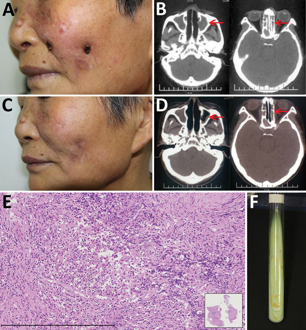
Mycobacterium gordonae infection in a 60-year-old immunocompetent woman, China. A) Facial lesions before treatment. Ulcers were erythematous and covered with yellow crusts. B) Computed tomography images before treatment show heterogeneous hypersignal in the ethmoid and left maxillary sinus (arrows). C) Facial lesions after treatment. Atrophic scars are seen at sites of previous lesions. D) Computed tomography images after treatment show recovery of the ethmoid sinus and left maxillary sinus (arr

Figure. Mycobacterium gordonae infection in a 60-year-old immunocompetent woman, China. A) Facial lesions before treatment. Ulcers were erythematous and covered with yellow crusts. B) Computed tomography images before treatment show heterogeneous hypersignal in the ethmoid and left maxillary sinus (arrows). C) Facial lesions after treatment. Atrophic scars are seen at sites of previous lesions. D) Computed tomography images after treatment show recovery of the ethmoid sinus and left maxillary sinus (arrows). E) Hematoxylin and eosin stain of nasal mucosa showing the infiltration of a large number of lymphocytes, a few histiocytes, and plasma cells. Scale bar corresponds to 400 µm. Inset shows the nasal mucosa sample (original magnification ×20). F) Tissue culture 3 weeks after incubation shows yolk-yellow bacteria growing in Löwenstein–Jensen medium. A color version of this figure is available online (https://wwwnc.cdc.gov/EID/article/23/7/16-2033-F1.htm).

Main Article

Page created: June 19, 2017
Page updated: June 19, 2017
Page reviewed: June 19, 2017
The conclusions, findings, and opinions expressed by authors contributing to this journal do not necessarily reflect the official position of the U.S. Department of Health and Human Services, the Public Health Service, the Centers for Disease Control and Prevention, or the authors' affiliated institutions. Use of trade names is for identification only and does not imply endorsement by any of the groups named above.
file_external