Volume 10, Number 12—December 2004
Research
Nonsusceptibility of Primate Cells to Taura Syndrome Virus
Carlos R. Pantoja* , Solangel A. Navarro*, Jaime Naranjo*, Donald V. Lightner*, and Charles P. Gerba*
Figure 5
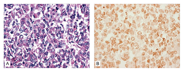
Figure 5. Absence of reaction by in situ hybridization (ISH) to the digoxigenin (DIG)-labeled Taura syndrome virus (TSV) probes within the BGMK cells harvested at day 7 postinjection with TSV. A) No cytopathic effect suggestive of TSV infection was evident by conventional hematoxylin/eosin-phloxin (H&E) histology (H&E stain; 100x). B) Consecutive histologic section to that shown in 4A, but subjected to ISH with DIG-labeled TSV probes specific for TSV. No reaction to TSV is apparent in the challenged cells (Bismarck Brown counterstain; 100x).
Page created: April 14, 2011
Page updated: April 14, 2011
Page reviewed: April 14, 2011
The conclusions, findings, and opinions expressed by authors contributing to this journal do not necessarily reflect the official position of the U.S. Department of Health and Human Services, the Public Health Service, the Centers for Disease Control and Prevention, or the authors' affiliated institutions. Use of trade names is for identification only and does not imply endorsement by any of the groups named above.