Volume 10, Number 6—June 2004
Research
Environmental Sources of Prion Transmission in Mule Deer
Cite This Article
Citation for Media
Abstract
Whether transmission of the chronic wasting disease (CWD) prion among cervids requires direct interaction with infected animals has been unclear. We report that CWD can be transmitted to susceptible animals indirectly, from environments contaminated by excreta or decomposed carcasses. Under experimental conditions, mule deer (Odocoileus hemionus) became infected in two of three paddocks containing naturally infected deer, in two of three paddocks where infected deer carcasses had decomposed in situ ≈1.8 years earlier, and in one of three paddocks where infected deer had last resided 2.2 years earlier. Indirect transmission and environmental persistence of infectious prions will complicate efforts to control CWD and perhaps other animal prion diseases.
Controlling and possibly eradicating animal prion diseases (1) are goals shared by the international community (2,3). However, progress toward eliminating prion diseases from food-producing animals worldwide has been hampered by incomplete knowledge about transmission and environmental persistence of these novel proteinaceous pathogens. Two prion diseases, scrapie of sheep and goats (4–8) and chronic wasting disease (CWD) of deer (Odocoileus spp.) and elk (Cervus elaphus nelsoni) (9–14), are particularly difficult to control because both are contagious among susceptible hosts. In contrast, bovine spongiform encephalopathy (BSE) does not appear to be contagious in cattle, but epidemics are sustained artificially through exposure to feed contaminated with infected bovine tissues (15); whether BSE in sheep is contagious remains undetermined (16). Both infected animals and environments apparently contaminated with the causative agent contribute to scrapie epidemics (4,6,8), and under some conditions, scrapie agents may persist in contaminated environments for years (7). Similarly, CWD is transmitted in the presence of infected mule deer (O. hemionus) (10), and circumstantial evidence exists for transmission from environments contaminated with the CWD agent (9,11,14). CWD epidemics do not appear to have been perpetuated by exposure to contaminated feed, but because ingestion of brain tissue can transmit CWD experimentally to deer (11,17), decomposed carcasses could serve as sources of infection in the environment.
Environmental sources of CWD infection represent potential obstacles to control in natural and captive settings. To investigate their role in transmission of this disease, we compared three potential sources of infection: infected live deer, decomposed infected deer carcasses, and an environment contaminated with residual excreta from infected deer.
We conducted a replicated experiment to compare CWD transmission from three infection sources: naturally infected captive mule deer (one infected deer/paddock), carcasses from naturally infected captive mule deer that had decomposed in situ ≈1.8 years earlier (one carcass/paddock), or undisturbed paddock environments where infected mule deer had last resided 2.2 years earlier. Each exposure source was replicated in three separate paddocks; two clean paddocks served as unexposed controls. Control paddocks and paddocks where live infected deer were added or where carcasses decomposed were constructed specifically for this experiment; these paddocks had never housed captive deer or elk and had been closed to access by free-ranging cervids for ≈17 years. Because clinical courses varied in naturally infected deer that served as sources of direct exposure, actual exposure periods varied from 0.75 year (replicate 3) to 1 year (replicate 1). Excreta-contaminated paddocks previously held 19 mule deer that had been orally inoculated during a 2-year pathogenesis study (11) that ended 2.2 years before our study began (≈3.8 infected deer x years of excreta/paddock, assuming equal distribution) but that had not held deer or elk in the interim. All three carcasses were from mule deer euthanized in end-stage clinical CWD. They had been left to decompose in intact form except for the removal of small pieces of brainstem used to confirm CWD infection; only the skeletal remains of carcasses were present at the start of the study.
Experimental animals included 31 free-ranging mule deer from two donor populations distant to endemic CWD foci. Experimental animals were captured from the grounds of the Rocky Mountain Arsenal National Wildlife Refuge (n = 17) and the U.S. Air Force Academy (n = 14), Colorado. We assumed that all experimental animals were free from CWD when they were introduced into the experiment, and surveillance data provided evidence that deer obtained from these herds were uninfected before exposure. Surveillance for CWD in the source populations (10,18) showed 0 positive cases in a sample of 210 adult deer from the refuge and 0 positive cases in a sample of 65 adult deer from the academy.
We used these data to estimate the probability that infection could have been caused by transmission from animals from the source herds. To do so, we estimated one-sided, exact 99% binomial confidence intervals (BCI) on the proportion of each population that could be positive for CWD (refuge = 0–0.022, academy = 0–0.068). We then used the upper limit of this interval to estimate the maximum prevalence,![]() , that could be reasonably expected in each of the source populations, given the inability to detect infections through surveillance. To assess whether observed results were likely due to preexisting infections, we treated each replicate (i.e., paddock) as an independent binomial experiment because the conditions in one paddock had no opportunity to influence the events in another paddock. Thus, for each replicate where infection occurred, we calculated the probability of at least one positive (i.e., “success”) given the number of animals introduced to that replicate from the source population (i.e., “trials”), on the assumption that the probability of drawing a positive from the source population was
, that could be reasonably expected in each of the source populations, given the inability to detect infections through surveillance. To assess whether observed results were likely due to preexisting infections, we treated each replicate (i.e., paddock) as an independent binomial experiment because the conditions in one paddock had no opportunity to influence the events in another paddock. Thus, for each replicate where infection occurred, we calculated the probability of at least one positive (i.e., “success”) given the number of animals introduced to that replicate from the source population (i.e., “trials”), on the assumption that the probability of drawing a positive from the source population was ![]() . When two replicates within an exposure category showed infections, we estimated the probability that cases in both replicates resulted from introducing infected animals (and not from our experiment) as the product of the individual replicate probabilities.
. When two replicates within an exposure category showed infections, we estimated the probability that cases in both replicates resulted from introducing infected animals (and not from our experiment) as the product of the individual replicate probabilities.
We captured deer during March and May 2002 and transported them to the Colorado Division of Wildlife’s Foothills Wildlife Research Facility, where they were confined in outdoor paddocks of ≈800 m2 (three replicate paddocks/exposure route, three deer/paddock); four deer were held in the two clean paddocks as unexposed controls. Each replicate of exposure paddocks was initially stocked with three mule deer. Shortly after arrival, one deer was moved to a different paddock within the same exposure condition to resolve social strife, and four fawns were born into three other paddocks; these changes are reflected in denominators in the Table. The distribution of prion protein genotype at codon 225 (serine [S]/phenylalanine [F] [19]) did not differ (Fisher exact test p = 0.6) among the four groups (three exposure groups + control).
Deer were fed alfalfa hay and a pelleted supplement; diets contained no animal protein or other animal byproducts. Individual paddocks and exposure blocks were physically segregated to prevent cross-transmission within and among exposure categories; dedicated clothing and equipment were used to minimize potential cross-contamination, but other potential fomites, like small mammals, birds, and insects, could not be controlled. However, transmission by routes such as these would be consistent with hypothesized transmission from environmental sources rather than direct animal-to-animal contact. After the animals had undergone ≈1 year of exposure to respective sources of infection, we obtained biopsied tonsil specimens from each participant deer and conducted an immunohistochemical analysis using anti-PrP MAb 99/97.6.1 (20,21). Upon detecting >1 infected deer in a paddock, we removed all inhabitants of that paddock and confirmed CWD infection in animals with positive biopsy results (20). Study protocols were reviewed and approved by the Colorado Division of Wildlife Animal Care and Use Committee
Mule deer exposed to contaminated environments or to infected deer contracted CWD (Table). None of the unexposed deer were infected. One or more introduced deer became infected in two of three paddocks containing a naturally infected deer, in two of three paddocks containing a decomposed deer carcass, and in one of three paddocks contaminated with residual deer excreta (Table) within 1 year of exposure. Infected deer included unrelated animals from both donor herds (2/17, 3/14; Fisher exact test p = 0.64), as well as one of four fawns born during the study. Males (4/16) and females (2/15) were infected at equivalent rates (Fisher exact test p = 0.65); similarly, deer of all three codon 225 genotypes (SS = 6/26, SF = 0/7, FF = 0/2) were infected at equivalent rates (Fisher exact test p = 0.52). Deer with positive biopsy results appeared healthy and did not show signs of CWD, consistent with early (<1 year in duration) infections (11,17).
On the basis of prior data from surveillance of source populations, our results were not likely explained by the null hypothesis of infections introduced from the source populations (p = 0.036 for academy source deer and p < 0.0001 for refuge source deer). The probability of prior infection accounting for our results in the pattern observed (Table) was p < 0.0013 for the infected animal exposure, p < 0.037 for the carcass exposure, p < 0.064 for the excreta exposure, and overall p ≈ 0.000003 for the observed results arising from preexisting infections. Because these probabilities were based on one-sided, upper 99% BCIs, we can conservatively reject the null hypothesis of infection arising from the source populations. The only remaining possibility is that infections arose from experimental exposures that included environments harboring the infectious agent from excreta or decomposed carcasses.
Figure
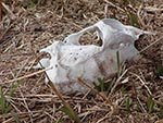
Figure. Green forage growing at the site where a deer carcass infected with chronic wasting disease had decomposed. Such sites were attractive to deer, as illustrated by the grass blades recently cropped...
Prions cannot be directly demonstrated in excreta or soil. However, CWD infection–specific protease-resistant prion protein (PrPCWD) accumulates in gut-associated lymphoid tissues (e.g., tonsils, Peyer patches, and mesenteric lymph nodes) of infected mule deer (11,17,22), which implicates alimentary shedding of the CWD agent in both feces and saliva (10,11,17). Because PrPCWD becomes progressively abundant in nervous system and lymphoid tissues through the disease course (11), carcasses of deer succumbing to CWD also likely harbor considerable infectivity and thus serve as foci of infection. We could not determine the precise mechanism for CWD transmission in excreta-contaminated paddocks, but foraging and soil consumption seemed most plausible. Deer did not actively consume decomposed carcass remains, but they did forage in the immediate vicinity of carcass sites where a likely nutrient flush (23) produced lush vegetation (Figure).
Our findings show that environmental sources of infectivity may contribute to CWD epidemics and illustrate the potential complexity of such epidemics in natural populations. The relative importance of different routes of infection from the environment cannot be discerned from our experiment, but each could play a role in sustaining natural epidemics. Although confinement likely exaggerated transmission probabilities, conditions simulated by this experiment do arise in the wild. Mule deer live in established home ranges and show strong fidelity to historic home ranges (24–26). As a result of such behavior, encounters with contaminated environments will occur more frequently than if deer movements were random. Feces and carcass remains are routinely encountered on native ranges, thus representing natural opportunities for exposure. Social behavior of deer, particularly their tendency to concentrate and become sedentary on their winter range, also may increase the probability of coming into contact with sources of infection in their environment.
The ability of the CWD agent to persist in contaminated environments for >2 years may further increase the probability of transmission and protract epidemic dynamics (8). Because infectivity in contaminated paddocks could not be measured, neither the initial levels nor degradation rate of the CWD agent in the environment was estimable. However, the observed persistence of the CWD agent was comparable to that of the scrapie agent, which persisted in paddocks for ≈1 to 3 years after removal of naturally infected sheep (7). Similarities between the CWD and scrapie agents suggest that environmental persistence may be a common trait of prions. Whether persistence of the BSE prion in contaminated feed production facilities or in environments where cattle reside contributed to BSE cases in the United Kingdom after feed bans were enacted (27) remains uncertain but merits further consideration.
Indirect transmission and environmental persistence of prions will complicate efforts to control CWD and perhaps other animal prion diseases. Historically, control strategies for animal prion diseases have focused on infected live animals as the primary source of infection. Although live deer and elk represent the most plausible mechanism for geographic spread of CWD, our data show that environmental sources could contribute to maintaining and prolonging local epidemics, even when all infected animals are eliminated. Moreover, the efficacy of various culling strategies as control measures depends in part on the rates at which the CWD agent is added to and lost from the environment. Consequently, these dynamics and their implications for disease management need to be more completely understood.
Dr. Miller is a wildlife veterinarian with the Wildlife Research Center, Colorado Division of Wildlife. His research has focused on the ecology and management of infectious diseases in free-ranging wildlife.
Acknowledgments
We thank T. Baker, T. Davis, P. Jeager, and others for assisting in field work, animal care, and laboratory aspects of this study; K. Cramer, J. Jewell, and M. Conner for providing genotype data and analyses; J. Hoeting, M. Samuel, and two anonymous reviewers for providing helpful comments on earlier drafts of our paper.
Our work was supported by the Colorado Division of Wildlife, the University of Wyoming, and National Science Foundation-National Institutes of Health Grant DEB-0091961.
References
- Vallat B. Preface. In: Lasmézas CI, Adams DB, editors. Risk analysis of prion diseases in animals. Rev Sci Tech. 2003;22:7–12.
- World Health Organization. Consultation on public health and animal transmissible spongiform encephalopathies: epidemiology, risk and research requirements (document WHO/CDS/CSR/APH/2000.2) [monograph on the Internet]. Geneva; 2000 [cited 2000 Sep 17]. Available from: http://www.who.int/emc‑documents/tse/whocdscsraph2002c.html
- Greig JR. Scrapie observations on the transmission of the disease by mediate contact. Vet J. 1940;96:203–6.
- Hoinville LJ. A review of the epidemiology of scrapie in sheep. Rev Sci Tech. 1996;15:827–52.PubMedGoogle Scholar
- Hourrigan J, Klingsporn A, Clark WW, de Camp M. Epidemiology of scrapie in the United States. In: Prusiner SB, Hadlow WJ, editors. Slow transmissible diseases of the nervous system: volume I. New York: Academic Press; 1979. p. 331−56.
- Pálsson PA. Rida (scrapie) in Iceland and its epidemiology. In: Prusiner SB, Hadlow WJ, editors. Slow transmissible diseases of the nervous system: volume I. New York: Academic Press; 1979. p. 357−66.
- Woolhouse MEJ, Stringer SM, Matthews L, Hunter N, Anderson RM. Epidemiology and control of scrapie within a sheep flock. Proc Biol Sci. 1998;265:1205–10. DOIPubMedGoogle Scholar
- Miller MW, Wild MA, Williams ES. Epidemiology of chronic wasting disease in Rocky Mountain elk. J Wildl Dis. 1998;34:532–8.PubMedGoogle Scholar
- Miller MW, Williams ES. Horizontal prion transmission in mule deer. Nature. 2003;425:35–6. DOIPubMedGoogle Scholar
- Williams ES, Miller MW. Chronic wasting disease in deer and elk in North America. In: Bengis RG, editor. Infectious diseases of wildlife: detection, diagnosis, and management, part two. Rev Sci Tech Off Int Epiz. 2002;21:305–16.
- Williams ES, Young S. Chronic wasting disease of captive mule deer: a spongiform encephalopathy. J Wildl Dis. 1980;16:89–98.PubMedGoogle Scholar
- Williams ES, Young S. Spongiform encephalopathy of Rocky Mountain elk. J Wildl Dis. 1982;18:465–71.PubMedGoogle Scholar
- Williams ES, Young S. Spongiform encephalopathies in Cervidae. Rev Sci Tech. 1992;11:551–67.PubMedGoogle Scholar
- Wilesmith JW, Ryan JBM, Atkinson MJ. Bovine spongiform encephalopathy: epidemiological studies of the origin. Vet Rec. 1991;128:199–203. DOIPubMedGoogle Scholar
- Kao RR, Gravenor MB, Baylis M, Bostock CJ, Chihota CM, Evans JC, The potential size and duration of an epidemic of bovine spongiform encephalopathy in British sheep. Science. 2002;295:332–5. DOIPubMedGoogle Scholar
- Sigurdson CJ, Williams ES, Miller MW, Spraker TR, O’Rourke KI, Hoover EA. Oral transmission and early lymphoid tropism of chronic wasting disease PrPres in mule deer fawns (Odocoileus hemionus). J Gen Virol. 1999;80:2757–64.PubMedGoogle Scholar
- Colorado Division of Wildlife. Prevalence and distribution of chronic wasting disease in Colorado 2002−2003 [monograph on the Internet]. 2003 Aug [cited 2003 Sep 4]. Available from: http://wildlife.state.co.us/cwd/pdf/prevalencesummary8-031.pdf
- Brayton KA, O’Rourke KI, Lyda AK, Miller MW, Knowles DP Jr. A processed pseudogene contributes to apparent mule deer prion gene heterogeneity. Gene. 2004;326:167–73. DOIPubMedGoogle Scholar
- Miller MW, Williams ES. Detecting PrPCWD in mule deer by immunohistochemistry of lymphoid tissues. Vet Rec. 2002;151:610–2. DOIPubMedGoogle Scholar
- Wolfe LL, Conner MM, Baker TH, Dreitz VJ, Burnham KP, Williams ES, Evaluation of antemortem sampling to estimate chronic wasting disease prevalence in free-ranging mule deer. J Wildl Manage. 2002;66:564–73. DOIGoogle Scholar
- Spraker TR, Zink RN, Cummings BA, Wild MA, Miller MW, O’Rourke KI. Comparison of histological lesions and immunohistochemical staining of protease resistant prion protein in a naturally occurring spongiform encephalopathy of free-ranging mule deer (Odocoileus hemionus) with those of chronic wasting disease of captive mule deer. Vet Pathol. 2002;39:110–9. DOIPubMedGoogle Scholar
- Towne EG. Prairie vegetation and soil nutrient responses to ungulate carcasses. Oecologia. 2000;122:232–9. DOIGoogle Scholar
- Conner MM, Miller MW. Movement patterns and spatial epidemiology of prion disease in mule deer population units. Ecol Appl. 2004;14. In press. DOIGoogle Scholar
- Kufeld RC, Bowden DC, Schrupp DL. Distribution and movements of female mule deer in the Rocky Mountain foothills. J Wildl Manage. 1989;53:871–7. DOIGoogle Scholar
- Mackie RJ. Mule deer habitat. In: Gerlach D, Atwater S, Schnell J, editors. Deer. Mechanicsburg (PA): Stackpole Books; 1994. p. 286−96.
- Wilesmith JW. Preliminary epidemiological analyses of the first 16 cases of BSE born after July 31, 1996, in Great Britain. Vet Rec. 2002;151:451–2. DOIPubMedGoogle Scholar
Figure
Table
Cite This ArticleTable of Contents – Volume 10, Number 6—June 2004
| EID Search Options |
|---|
|
|
|
|
|
|
Please use the form below to submit correspondence to the authors or contact them at the following address:
Michael W. Miller, Colorado Division of Wildlife, Wildlife Research Center, 317 West Prospect Road, Fort Collins, CO 80526-2097, USA; fax: 970-472-4457
Top