Volume 11, Number 12—December 2005
Letter
Echinococcus multilocularis in Estonia
Figure
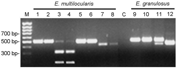
Figure. Diagnostic polymerase chain reaction (PCR) restriction fragment length polymorphism analysis for Echinococcus multilocularis (lanes 1–8, 2 specimens in parallel) and E. granulosus (lanes 9–12, 1 specimen). Lane M: Gene Ruler 100-bp DNA ladder; lane C: negative control without DNA; lanes 1 and 2: amplification of E. multilocularis DNA with Eg9 PCR; lanes 3 and 4: amplification of E. multilocularis DNA with Eg9 PCR, followed by cleavage with enzyme CfoI; lanes 5 and 6: amplification of E. multilocularis DNA with Eg9 PCR, followed by cleavage with enzyme RsaI; lanes 7 and 8: amplification of E. multilocularis DNA with Eg16 PCR; lane 9: amplification of E. granulosus DNA with Eg9 PCR; lane 10: amplification of E. granulosus DNA with Eg9 PCR, followed by cleavage with enzyme CfoI; lane 11: amplification of E. granulosus DNA with Eg9 PCR, followed by cleavage with enzyme RsaI; lane 12: amplification of E. granulosus DNA with Eg16 PCR.