Skip directly to site content Skip directly to page options Skip directly to A-Z link Skip directly to A-Z link Skip directly to A-Z link
Volume 11, Number 12—December 2005
Dispatch

Human Rickettsia felis Infection, Canary Islands, Spain

Jose-Luis Pérez-Arellano*†1, Florence Fenollar‡1, Alfonso Angel-Moreno*†, Margarita Bolaños*†, Michele Hernández*†, Evora Santana*†, Marion Hemmersbach-Miller*†, Antonio-M Martín*†, and Didier Raoult‡Comments to Author 
Author affiliations: *Hospital Universitario Insular de Las Palmas, Canary Islands, Spain; †Universidad de Las Palmas de Gran Canaria, Canary Islands, Spain; ‡Université de la Mediterranée, Marseille, France

Main Article

Figure

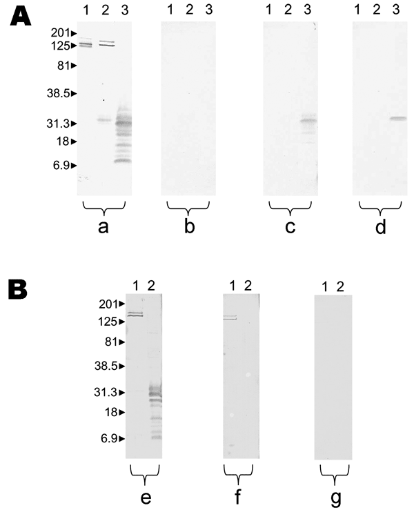
Results of Western blot performed with serum samples from patient 5 with Rickettsia felis infection and patient 10 with R. typhi infection. Molecular masses (in kilodaltons) are given to the left of panels. A) Patient with R. felis infection; a, untreated serum analyzed by using R. conorii (lane 1), R. typhi (lane 2), and R. felis (lane 3); b, R. felis–adsorbed serum analyzed by using R. conorii (lane 1), R. typhi (lane 2), R. felis (lane 3); all antibodies were removed; c, R. typhi–adsorbed ser

Figure. Results of Western blot performed with serum samples from patient 5 with Rickettsia felis infection and patient 10 with R. typhi infection. Molecular masses (in kilodaltons) are given to the left of panels. A) Patient with R. felis infection; a, untreated serum analyzed by using R. conorii (lane 1), R. typhi (lane 2), and R. felis (lane 3); b, R. felis–adsorbed serum analyzed by using R. conorii (lane 1), R. typhi (lane 2), R. felis (lane 3); all antibodies were removed; c, R. typhi–adsorbed serum analyzed by using R. typhi (lane 1) and R. felis (lane 2); antibodies to R. felis remained; d, R. conorii–adsorbed serum analyzed by using R. conorii (lane 1), R. typhi (lane 2), R. felis (lane 3); antibodies to R. felis remained. B) Patient with murine typhus; e, untreated serum analyzed by using R. typhi (lane 1) and R. felis (lane 2); f, R. felis–adsorbed serum analyzed by using R. typhi (lane 1) and R. felis (lane 2); antibodies to R. typhi remained; g, R. typhi–adsorbed serum analyzed by using R. typhi (lane 1) and R. felis (lane 2); all antibodies were removed.

Main Article

1These authors contributed equally to this article.

Page created: February 02, 2012
Page updated: February 02, 2012
Page reviewed: February 02, 2012
The conclusions, findings, and opinions expressed by authors contributing to this journal do not necessarily reflect the official position of the U.S. Department of Health and Human Services, the Public Health Service, the Centers for Disease Control and Prevention, or the authors' affiliated institutions. Use of trade names is for identification only and does not imply endorsement by any of the groups named above.
file_external