Volume 11, Number 5—May 2005
Research
Venezuelan Equine Encephalitis Virus Infection of Spiny Rats
Anne-Sophie Carrara*, Marta Gonzales†, Cristina Ferro†, Margarita Tamayo†, Judith Aronson*, Slobodan Paessler*, Michael Anishchenko*, Jorge Boshell†, and Scott C. Weaver*
Figure 3
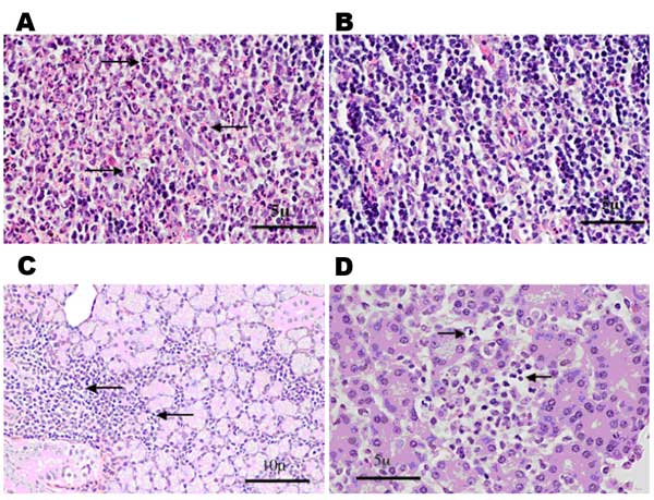
Figure 3. . Histologic staining (hematoxylin and eosin) of spiny rat lymph nodes (A and B), salivary glands (C), and pancreas (D) after subcutaneous inoculation of 3 log10 PFU of Venezuelan equine encephalitis virus strain Co97-0054. A) Popliteal draining lymph node 24 h postinfection, showing the presence of a polymorphonuclear leukocyte infiltrate (arrows). B) Contralateral popliteal lymph node 24 hr postinfection from same animal. No proinfiltration was visible. C) Chronic inflammation of the salivary gland (arrows) day 3 postinfection. D) Asenea with focal necrosis (arrows) day 3 postinfection. (Magnification ×40.)
Page created: April 24, 2012
Page updated: April 24, 2012
Page reviewed: April 24, 2012
The conclusions, findings, and opinions expressed by authors contributing to this journal do not necessarily reflect the official position of the U.S. Department of Health and Human Services, the Public Health Service, the Centers for Disease Control and Prevention, or the authors' affiliated institutions. Use of trade names is for identification only and does not imply endorsement by any of the groups named above.