Volume 11, Number 8—August 2005
Perspective
Virology, Pathology, and Clinical Manifestations of West Nile Virus Disease
Edward B. Hayes* , James J. Sejvar†, Sherif R. Zaki†, Robert S. Lanciotti*, Amy V. Bode*, and Grant L. Campbell*
Figure
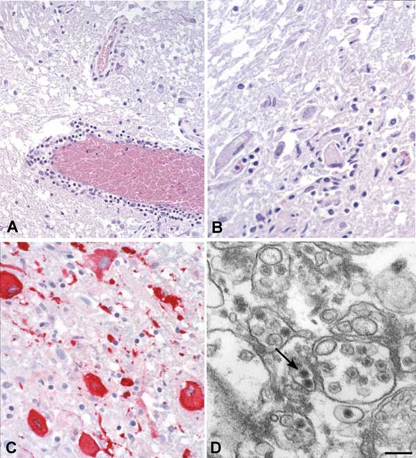
Figure. . Histopathologic features of West Nile virus (WNV) in human tissues. Panels A and B show inflammation, microglial nodules, and variable necrosis that occur during WNV encephalitis; panel C shows WNV antigen (red) in neurons and neuronal processes using an immunohistochemical stain; panel D is an electron micrograph of WNV in the endoplasmic reticulum of a nerve cell (arrow). Bar = 100 nm.
Page created: April 23, 2012
Page updated: April 23, 2012
Page reviewed: April 23, 2012
The conclusions, findings, and opinions expressed by authors contributing to this journal do not necessarily reflect the official position of the U.S. Department of Health and Human Services, the Public Health Service, the Centers for Disease Control and Prevention, or the authors' affiliated institutions. Use of trade names is for identification only and does not imply endorsement by any of the groups named above.