Volume 12, Number 1—January 2006
Dispatch
Rickettsia felis Infection, Tunisia
Abir Znazen*†, Jean-Marc Rolain*, Nader Hammami†, Adnane Hammami†, Mounir Ben Jemaa‡, and Didier Raoult*
Figure
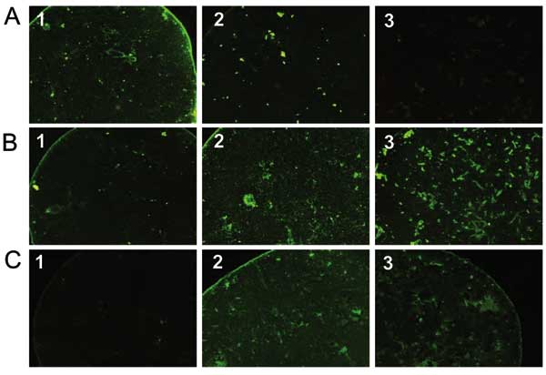
Figure. Pictures of immunofluorescence assay performed on serum specimens with proven Rickettsia conorii (A), R. felis (B), or R. typhi (C) infection showing cross-reactive antibodies. Antigens tested were R. conorii (column 1), R. felis (column 2), and R. typhi (column 3). The serum with R. conorii infection reacts with R. conorii and R. felis antigens but not with R. typhi (A). Conversely, the serum with R. typhi infection reacts with R. typhi and R. felis but not with R. conorii (C). Finally, the serum with R. felis infection reacts with R. felis, R. conorii, and R. typhi.
Page created: February 16, 2012
Page updated: February 16, 2012
Page reviewed: February 16, 2012
The conclusions, findings, and opinions expressed by authors contributing to this journal do not necessarily reflect the official position of the U.S. Department of Health and Human Services, the Public Health Service, the Centers for Disease Control and Prevention, or the authors' affiliated institutions. Use of trade names is for identification only and does not imply endorsement by any of the groups named above.