Volume 12, Number 11—November 2006
Research
Susceptibility of North American Ducks and Gulls to H5N1 Highly Pathogenic Avian Influenza Viruses
Figure 2
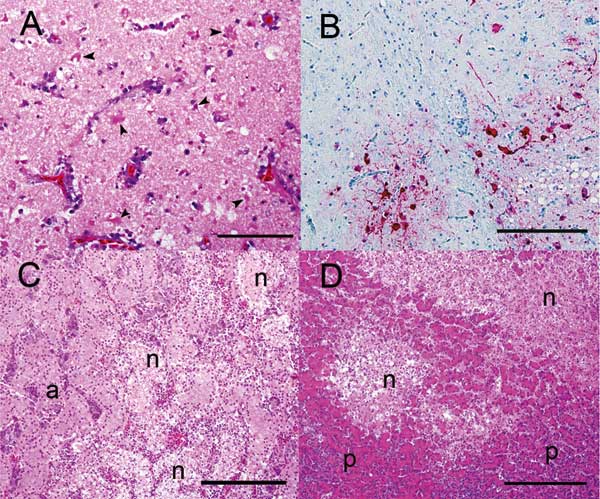
Figure 2. Photomicrographs of visceral organs from a wood duck that died after intranasal inoculation with a highly pathogenic avian influenza H5N1 virus. A) Brain with severe, multifocal to coalescing neuronal necrosis. Note the numerous necrotic neurons (arrowheads). Hematoxylin and eosin (HE) stain; bar =100 μm. B) Brain. Note the viral antigen (red) detected in the nucleus of several neurons. The unaffected brain tissue is blue. Immunohistochemical stain with hematoxylin counterstain; bar = 200 μm. C) Adrenal gland with necrotizing adrenalitis. Note the multiple foci of necrosis (n) surrounded by normal adrenal parenchyma (a). HE stain; bar = 200 μm. D) Pancreas with necrotizing pancreatitis. Note the 2 well-demarcated areas of necrosis (n) within the normal pancreatic tissue (p).