Volume 12, Number 12—December 2006
Research
Human Metapneumovirus in Turkey Poults
Figure 2
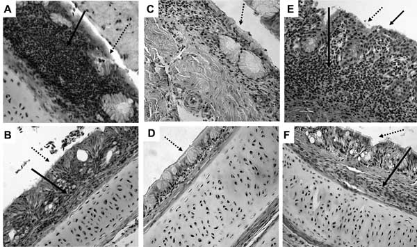
Figure 2. Histopathologic appearance of nasal turbinate and trachea tissue (magnification ×200). A) Nasal turbinates of turkey poults exposed to human metapneumovirus (hMPV) B2, showing infiltration of inflammatory cells (hematoxylin and eosin staining; solid arrow ). B) Trachea of turkey poults exposed to hMPV B2, showing mild inflammation with infiltration of a few inflammatory cells in the lamina propria (solid arrow). C) Nasal turbinate of sham-inoculated turkey poults. D) Trachea of sham-inoculated turkey poults. E) Nasal turbinate of turkey poults exposed to avian metapneumovirus (aMPV C), showing infiltration of inflammatory cells and multifocal loss of cilia (solid arrows). F) Trachea of turkey poults exposed to aMPV C, showing mild inflammation with infiltration of a few inflammatory cells in the lamina propria (solid arrow). Dotted arrows indicate mucosal surface.