Volume 12, Number 2—February 2006
Research
Helicobacter pullorum in Chickens, Belgium
Cite This Article
Citation for Media
Abstract
A total of 110 broilers from 11 flocks were tested for Helicobacter pullorum by polymerase chain reaction; positive samples were reexamined with a conventional isolation method. H. pullorum isolates were examined by amplified fragment length polymorphism (AFLP) fingerprinting for interstrain genetic diversity and relatedness. Sixteen isolates from cecal samples from 2 different flocks were obtained. AFLP analysis showed that these isolates and 4 additional isolates from a different flock clustered according to their origin, which indicates that H. pullorum colonization may occur with a single strain that disseminates throughout the flock. Strains isolated from different hosts or geographic sources displayed a distinctive pattern. H. pullorum is present in approximately one third of live chickens in Belgium and may represent a risk to human health.
Helicobacter pullorum was originally isolated from the feces and damaged livers of broilers and laying hens (1,2). It was defined as a new species in 1994 by Stanley et al. (1). H. pullorum is a gram-negative, slightly curved rod with monopolar, nonsheathed flagella. It is bile resistant and requires a microaerobic environment supplemented with H2 in which growth occurs at 37°C and 42°C (1,3–6). Enterohepatic Helicobacter species, including H. pullorum, are increasingly recognized as microbial pathogens in humans and animals (3,5,7–9). H. pullorum has been linked with enteritis and hepatitis in broiler chickens and laying hens and diarrhea, gastroenteritis, and liver disease in humans (1,2,5–8,10,11). H. pullorum can contaminate poultry carcasses at the abattoir and can be considered a foodborne human pathogen (4,8,12).
Almost no data are available on the prevalence of this species in poultry. Research that could generate these data is hampered by the fastidious growth requirements of H. pullorum and the phenotypic similarity between member species of the genera Helicobacter and Campylobacter (3,4,12). H. pullorum in chickens has been studied on only 2 occasions when the organism was detected by using isolation (4,7). Furthermore, no valid epidemiologic research methods have been recommended.
This study's objective was to determine the occurrence of H. pullorum in broilers by using both polymerase chain reaction (PCR) and isolation. In addition, amplified fragment length polymorphism profiling (AFLP) was conducted to investigate the genetic relatedness between H. pullorum isolates.
Sample Origin
Samples from the gastrointestinal tracts and livers of 110 broiler chickens, 10 per flock (flock number 1–11), collected at a poultry abattoir, were studied. Each gastrointestinal tract and liver sample was deposited in a separate waterproof plastic bag. Samples were taken from the liver, cecum, jejunum, and colon for PCR and isolation within 3 hours after collection. All samples were stored at –20°C and –70°C for PCR and isolation, respectively, until further analysis, as described below.
Sample Processing
PCR and Gel Electrophoresis
DNA was extracted from ≈25 mg cecum, colon, jejunum, and liver tissue with a commercial tissue kit (DNeasy Tissue Kit, Qiagen, Venlo, the Netherlands). A PCR assay amplifying a 447-bp fragment of the 16S rRNA gene of H. pullorum was then used for detection purposes (1). From each sample, 2 μL template was added to 8 μL PCR mixture containing 0.03 U/μL Taq polymerase Platinum (Invitrogen Life Technologies, Merelbeke, Belgium), 10× PCR Buffer (Invitrogen Life Technologies), 3 mmol MgCl2 (Invitrogen, Life Technologies), 40 μmol/L each of deoxynucleoside triphosphate (Invitrogen Life Technologies), a final primer concentration of 0.5 μmol/L, and sterile distilled water. The conditions used for the amplifications were the following: an initial denaturation at 94°C for 5 min, followed by 35 cycles of denaturation at 94°C for 1 min, annealing at 60°C for 90 s, elongation at 72°C for 90 s, and a final elongation at 72°C for 5 min.
Five microliters of the PCR products of each sample were mixed with 3 μL of sample buffer 5× (50% glycerol, 1 mmol cresol red) and were subjected to electrophoresis through an agarose gel containing 1.5% Multi Purpose agarose (Boehringer, Mannheim, Germany) and 50 ng ethidium bromide in per milliliter 1× Tris-acetate ethylenediaminetetraacetic acid buffer (Amresco, Solon, OH, USA), pH 8. As molecular size marker, the Gene Ruler 100-bp DNA ladder plus (MBI Fermentas, St. Leon-Rot, Germany) was used. Electrophoresis was implemented at a constant voltage of 170 V in 0.5× Tris-acetate ethylenediaminetetraacetic buffer for 75 min. The gels were visualized by using the Image Master VDS (Pharmacia Biotech, Puurs, Belgium).
Isolation of H. pullorum
Recovery of H. pullorum isolates was attempted on all positive samples in the PCR analysis described above. The samples (200 mg) for isolation of H. pullorum were placed in a 1.5-mL tube with 400 μL of a mixture of 7.5 g glucose, 25 mL brain heart infusion broth (Oxoid, Basingstoke, England), and 75 mL sterile inactivated horse serum, and then homogenized. The various isolates were inoculated on brain heart infusion agar that was supplemented with 10% horse blood, amphotericin B 20 μg/mL (Fungizone, Bristol-Myers Squibb, Epernon, France), and Vitox (Oxoid) (blood agar). A modified filter technique of Steele and McDermott (13) was then used. Briefly, a sterile cellulose acetate membrane filter (0.45 μm) was applied with a sterile pair of tweezers directly onto the surface of the agar. When the filter was totally absorbed on the agar, ≈300 μL of the mixture was placed in the middle of the filter. After at least 1 hour of incubation at 37°C and 5% CO2, the filter was removed with a sterile pair of tweezers and the filtrate was streaked on the agar with a loop. Incubation was conducted in microaerobic conditions (5% H2, 5% CO2, 5% O2, and 85% N2) at 37°C for a minimum of 3 days. Very small, gray-white, hemolytic colonies were selected and purified on a blood agar plate. The colonial form and phenotypic characteristics (gram-negative, slightly curved rod, catalase and oxidase positive, and indoxyl acetate negative) of the isolates were used for presumptive identification. Confirmation was based on PCR and sequencing of a 447-bp fragment of the 16S ribosomal RNA gene, as described below.
Analysis of Nucleotide Sequences
The PCR product of the retrieved H. pullorum isolates was purified with the Qiaquick purification kit (Qiagen) and sequenced by using the same primers applied in the assay with the BigDye Terminator cycle sequencing kit (Applied Biosystems, Lennik, Belgium). Sequencing products were run on the ABI prism 3100 Genetic Analyzer (Applied Biosystems) by using 50-cm capillaries filled with Performance-Optimized-Polymer 6. The electrophoregrams were exported and converted to the Kodon software package (Applied Maths, Sint-Martems-Latem, Belgium). Sequences were compared to published H. pullorum 16S rRNA sequences obtained from GenBank (accession nos. AY631956, L36143, and L36144) by using BLAST software (available from http://www.ncbi.nlm.nih.gov/blast/).
AFLP
Twenty-two poultry and 3 human isolates were fingerprinted by using AFLP (Table 1). These included 16 isolates from flock numbers 5 and 9 screened in this study. In addition, 4 samples previously isolated from broilers' cecal droppings and the boots from another flock's farmer, 4 reference strains (2 of chicken and 2 of human), and 1 human strain isolated from diarrheic stool in our laboratory were included for comparison.
Restriction Endonuclease Digestion and Ligation of Adaptors for AFLP
DNA of H. pullorum isolates was extracted by using a commercial tissue kit (DNeasy Tissue Kit, Qiagen). An aliquot containing 200 ng DNA, determined by optic density (260/280 nm) measurement by using the Spectra Fluor (TECAN, Grödig, Salzburg, Austria), was digested for 2 h at 37°C with BglII (10U/μL) and Csp6I (10U/μL) (MBI Fermentas) in TAC-buffer as described by Vos et al. (14). Five microliters of DNA digest was used in a ligation reaction containing 130 μg/mL BglII adaptor-oligonucleotide and 13 μg/mL Csp6I adaptor-oligonucleotide (Invitrogen) (14), 10× T4 DNA ligase buffer, T4 DNA ligase (1 U/μL) (Amersham Pharmacia), and TAC-buffer in a final volume of 20 μL. After incubation for 2 h at 25°C, the 20 μL ligation reaction was diluted 25 times.
Direct Selective PCR Amplification of Diluted Ligation
Five microliters of the diluted ligation reaction were applied in the PCR assay. The primers used in this assay were BGL2F-0, 5´-GAG TAC ACT GTC GAT CT-3´ (FAM labeled, 5´-end) and CSP6I-A, 5´-GAG CTC TCC AGT ACT ACA-3´ (15). The PCR conditions were as follows: an initial denaturation at 94°C for 3 min; 35 cycles of denaturation at 94°C for 1 min, annealing at 54°C for 1 min, and elongation at 72°C for 90 s; and a final elongation at 72°C for 10 min.
Capillary Electrophoresis
PCR products were run on the ABI prism 3100 Genetic Analyzer (Applied Biosystems) by using the Fragile X Rox-1000 size standard and 50-cm capillaries filled with Performance-Optimized-Polymer 6. Electropherograms were analyzed with Genemapper U 3.5 Software (Applied Biosystems).
Numerical Analyses of AFLP Profiles
The program BioNumerics version 2.5 (Applied Maths) was used to perform numerical analyses of AFLP profiles. Strain relationships were inferred by use of the Pearson product-moment correlation coefficient and unweighted pair-group with mathematical average (UPGMA) clustering and depicted in a dendrogram (16).
PCR
In Table 2, the number of H. pullorum DNA–positive samples originating from the intestinal tract and liver is shown. In 4 flocks, all samples were negative for H. pullorum. In the other 7 flocks, positive samples were found. In the cecum and colon, a PCR reaction for H. pullorum was positive in 33.6% and 31.8% of the samples, respectively. In total, 10.9% of jejunum and 4.6% of liver samples were positive for H. pullorum.
Isolation of H. pullorum
Eight H. pullorum cecum isolates from flock number 5 and 8 H. pullorum cecum isolates from flock number 9 were obtained. The sequences of the amplified 447-bp fragment of the H. pullorum 16S ribosomal RNA gene isolates showed a similarity of 98%–100% to those from GenBank (accession nos. AY631956, L36142, and L36143).
AFLP
Figure
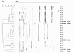
Figure. Chicken isolates and human strains of Helicobacter pullorum by amplified fragment length polymorphism.
AFLP analysis showed that isolates from each of the individual flocks examined clustered according to their flock of origin. The remaining chicken isolates and human strains each displayed a unique profile (Figure).
This study shows that H. pullorum is present in 33.6% of the cecal samples of broiler chickens collected at a poultry slaughterhouse during evisceration by using PCR. This microorganism was found in 7 of 11 flocks; 4 flocks were negative. Burnens et al. found a prevalence rate of 4% upon sampling cecal contents of broilers (7). The organism was detected by isolation. Considering the fastidious nature of this organism, this finding could explain this markedly lower percentage of positive birds. Additionally, in our study, cecal tissue, rather than cecal contents, was examined for the organism. Microorganisms related to H. pullorum adhere closely to the mucosa of the gastrointestinal tract. The phylogenetically related microorganism, C. jejuni, may tightly adhere to the brush borders of the intestine in chickens (17,18). The same phenomenon has also been documented for H. pylori in the stomach (19).
Comparing our study results to those obtained by Atabay et al. (4), the latter group found a higher occurrence of H. pullorum (60%) on poultry carcasses. This apparent discrepancy could be due to cross-contamination with cecal contents on the surface of broiler carcasses during poultry processing (4,8). Furthermore, contamination of the chicken body surface may occur during transportation to the abattoir. Fecal excretion of Campylobacter spp. may be increased because of stress during transportation and consequently may contaminate carcasses (20).
H. pullorum DNA was detected in only 5 (4.6%) liver and 11 (10.9%) jejunal samples, as opposed to 35 (31.8%) colonic and 37 (33.6%) cecal samples. Hence, one may assume that the lower segments of the intestinal tract are the predominant colonization sites for H. pullorum in broiler chickens. H. pullorum may gain access to the liver by retrograde transfer from the duodenum. Alternatively, it may translocate from the gut lumen to the portal circulation.
H. pullorum has been associated with vibrionic hepatitis in laying hens, both macroscopically and microscopically (7). In our study, no gross pathologic lesions were seen in the livers during sampling (data not shown).
Our modest isolation rate of H. pullorum from cecal samples may have been the result of examining frozen, as opposed to fresh, samples. However, we successfully recovered 16 isolates from 2 flocks, allowing (for the first time, to our knowledge) some analysis of the etiology of H. pullorum in broiler flocks to be undertaken. We used AFLP profiling for this purpose, a highly discriminatory method that has been successfully applied to molecular epidemiologic studies of several related species, including H. pylori (21,22), Arcobacter spp (15), and Campylobacter spp (23,24). Isolates from each of the individual flocks clustered according to their flock of origin, indicating a clonal relationship. In contrast, field and reference strains isolated from different hosts or geographic sources displayed a distinctive pattern. These data suggest that AFLP profiling has considerable potential for molecular epidemiologic studies of H. pullorum for the noted related species.
Several authors have suggested that H. pullorum has zoonotic potential and is involved in the pathogenesis of diarrhea and chronic liver diseases in humans (2,8,10,11). Retail raw poultry meats and other poultry products may constitute vehicles for human H. pullorum infections through carcass contamination, as previously reported for Arcobacter and Campylobacter species (8,25–27). Concerning health monitoring, PCR may be helpful in detecting this pathogen not only in intestinal tissue but also in broiler chicken cecal droppings.
In conclusion, this study shows that H. pullorum is a frequent intestinal colonizer of broiler chickens. PCR and isolation are useful tools to detect the species in intestinal tissue and in cecal droppings. AFLP profiling appears to be useful for molecular epidemiologic studies of this species.
Ms Ceelen is a veterinary PhD student at Ghent University in Belgium where the work described in this study was performed. Her research interests include bacterial pathogenesis and host-pathogen interactions, with a focus on Helicobacter spp.
Acknowledgments
We thank the abattoir Nollens for providing intestinal tracts and livers from poultry and Marc Heyndrickx for providing H. pullorum strains. We thank Jurgen De Craene for excellent technical assistance and Peter Dawyndt for his assistance in data analysis of the AFLP profiles.
This work was supported by a PhD grant from the Institute for the Promotion of Innovation by Science and Technology in Flanders (I.W.T. Vlaanderen) to Liesbeth Ceelen.
References
- Stanley J, Linton D, Burnens AP, Dewhirst FE, On SLW, Porter A, Helicobacter pullorum sp. nov.-genotype and phenotype of a new species isolated from poultry and from human patients with gastroenteritis. Microbiol. 1994;140:3441–9. DOIPubMedGoogle Scholar
- Burnens AP, Stanley J, Morgenstern R, Nicolet J. Gastroenteritis associated with Helicobacter pullorum. Lancet. 1994;344:1569–70. DOIPubMedGoogle Scholar
- On SLW, Holmes B, Sackin MJ. A probability matrix for the identification of campylobacters, helicobacters and allied taxa. J Appl Bacteriol. 1996;81:425–32.PubMedGoogle Scholar
- Atabay HI, Corry JEL, On SLW. Identification of unusual Campylobacter-like isolates from poultry products as Helicobacter pullorum. J Appl Microbiol. 1998;84:1017–24. DOIPubMedGoogle Scholar
- Fox JG. The expanding genus of Helicobacter: pathogenic and zoonotic potential. Semin Gastrointest Dis. 1997;8:124–41.PubMedGoogle Scholar
- Steinbrueckner B, Hearter G, Pelz K, Weiner S, Rump JA, Deissler W, Isolation of Helicobacter pullorum from patients with enteritis. Scand J Infect Dis. 1997;29:315–8. DOIPubMedGoogle Scholar
- Burnens AP, Stanley J, Nicolet J. Possible association of Helicobacter pullorum with lesions of vibrionic hepatitis in poultry. In: Newell DG, Ketley JM, and Feldman RA, editors. Campylobacters, helicobacters and related organisms. New York: Plenum Press; 1996.
- Fox JG, Dewhirst FE, Shen Z, Feng Y, Taylor NS, Paster BJ, Hepatic Helicobacter species identified in bile and gallbladder tissue from Chileans with chronic cholecystitis. Gastroenterology. 1998;114:755–63. DOIPubMedGoogle Scholar
- On SLW, Hynest S, Wadström T. Extragastric Helicobacter species. Helicobacter. 2002;7:S63–7. DOIPubMedGoogle Scholar
- Young VB, Chien CC, Knox KA, Taylor NS, Schauer DB, Fox JG. Cytolethal distending toxin in avian and human isolates of Helicobacter pullorum. J Infect Dis. 2000;182:620–3. DOIPubMedGoogle Scholar
- Ceelen L, Decostere A, Verschraegen G, Ducatelle R, Haesebrouck F. Prevalence of Helicobacter pullorum among patients with gastrointestinal disease and clinically healthy persons. J Clin Microbiol. 2005;43:2984–6. DOIPubMedGoogle Scholar
- Gibson JR, Ferrus MA, Woodward D, Xerry J, Owen RJ. Genetic diversity in Helicobacter pullorum and poultry sources identified by an amplified fragment length polymorphism technique and pulsed-field gel electrophoresis. J Appl Microbiol. 1999;87:602–10. DOIPubMedGoogle Scholar
- Steele TW, McDermott SN. The use of membrane filters applied directly to the surface of agar plates for the isolation of Campylobacter jejuni from faeces. Pathology. 1984;16:263–5. DOIPubMedGoogle Scholar
- Vos P, Hogers R, Bleeker M, Reijans M, van de Lee T, Hornes M, AFLP: a new technique for DNA fingerprinting. Nucleic Acids Res. 1995;11:4407–14. DOIPubMedGoogle Scholar
- Kokotovic B, On SLW. High-resolution genomic fingerprinting of Campylobacter jejuni and Campylobacter coli by analysis of amplified fragment length polymorphisms. FEMS Microbiol Lett. 1999;173:77–84. DOIPubMedGoogle Scholar
- On SL, Harrington CS, Atabay HI. Differentiation of Arcobacter species by numerical analysis of AFLP profiles and description of a novel Arcobacter from pig abortions and turkey faeces. J Appl Microbiol. 2003;95:1096–105. DOIPubMedGoogle Scholar
- Sanyal SC, Islam KM, Neogy PK, Islam M, Speelman P, Huq MI. Campylobacter jejuni diarrhea model in infant chickens. Infect Immun. 1984;43:931–6.PubMedGoogle Scholar
- Ruiz-Palacios GM, Escamilla E, Torres N. Experimental Campylobacter diarrhea in chickens. Infect Immun. 1981;34:250–5.PubMedGoogle Scholar
- Clyne M, Drumm B. Adherence of Helicobacter pylori to primary human gastrointestinal cells. Infect Immun. 1993;61:4051–7.PubMedGoogle Scholar
- Whyte P, Collins JD, McGill K, Monahan C, O'Mahony H. The effect of transportation stress on excretion rates of campylobacters in market-age broilers. Poult Sci. 2001;80:817–20.PubMedGoogle Scholar
- Fox JG. The expanding genus of Helicobacter: pathogenic and zoonotic potential. Semin Gastrointest Dis. 1997;8:124–41.PubMedGoogle Scholar
- Ananieva O, Nilsson I, Vorobjovat T, Uibo R, Wadstrom T. Immune responses to bile-tolerant Helicobacter species in patients with chronic liver diseases, a randomized population group, and healthy blood donors. Clin Diagn Lab Immunol. 2002;9:1160–4.PubMedGoogle Scholar
- Siemer BL, Harrington CS, Nielsen EM, Borck B, Nielsen NL, Engberg J, Genetic relatedness among Campylobacter jejuni serotyped isolates of diverse origin as determined by numerical analysis of amplified fragment length polymorphism (AFLP) profiles. J Appl Microbiol. 2004;96:795–802. DOIPubMedGoogle Scholar
- Siemer BL, Nielsen EM, On SLW. Identification and molecular epidemiology of Campylobacter coli isolates from human gastroenteritis, food and animal sources evaluated by amplified fragment length (AFLP) analysis and Penner serotyping. Appl Environ Microbiol. 2005;71:1953–8. DOIPubMedGoogle Scholar
- Houf K, Tutenel A, De Zutter L, Van Hoof J, Vandamme P. Development of a multiplex PCR assay for the simultaneous detection and identification of Arcobacter butzleri, Arcobacter cryaerophilus and Arcobacter skirrowii. FEMS Microbiol Lett. 2000;193:89–94. DOIPubMedGoogle Scholar
- Houf K, Devriese LA, De Zutter L, Van Hoof J, Vandamme P. Development of a new protocol for the isolation and quantification of Arcobacter species from poultry products. Int J Food Microbiol. 2001;71:189–96. DOIPubMedGoogle Scholar
- Antolin A, Gonzalez I, Garcia T, Hernandez PE, Martin R. Arcobacter spp. enumeration in poultry meat using a combined PCR-ELISA assay. Meat Sci. 2001;59:169–74. DOIPubMedGoogle Scholar
Figure
Tables
Cite This ArticleTable of Contents – Volume 12, Number 2—February 2006
| EID Search Options |
|---|
|
|
|
|
|
|
Please use the form below to submit correspondence to the authors or contact them at the following address:
Liesbeth M. Ceelen, Department of Pathology, Bacteriology and Avian Diseases, Faculty of Veterinary Medicine, Ghent University, Salisburylaan 133, 9820 Merelbeke, Belgium; fax: 32-9-264-74-94
Top