Volume 12, Number 3—March 2006
Research
Pneumonic Plague Cluster, Uganda, 2004
Elizabeth M. Begier* , Gershim Asiki†, Zaccheus Anywaine†, Brook Yockey‡, Martin Schriefer‡, Philliam Aleti§, Asaph Ogen-Odoi§, J. Erin Staples*‡, Christopher Sexton‡, Scott Bearden‡, and Jacob L. Kool‡
Figure 1
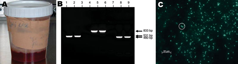
Figure 1. A) Grossly bloody sputum sample obtained from the surviving patient (caregiver B2) 30 h after onset of primary pneumonic plague. B) Polymerase chain reaction results of sputum sample from caregiver B2. Lanes 1–3, caf1; lanes 4–6, repA1; lanes 7–9, pla. Lanes 1, 4, and 7 are positive controls; lanes 2, 5, and 8 are patient samples; lanes 3, 6, and 9 are negative controls. C) Anti-F1 direct fluorescent antibody staining of sputum sample from caregiver B2. Numerous bacteria with classic halo structures are characteristic of Yersinia pestis. The circled bacterium classically depicts this halo.
Page created: January 27, 2012
Page updated: January 27, 2012
Page reviewed: January 27, 2012
The conclusions, findings, and opinions expressed by authors contributing to this journal do not necessarily reflect the official position of the U.S. Department of Health and Human Services, the Public Health Service, the Centers for Disease Control and Prevention, or the authors' affiliated institutions. Use of trade names is for identification only and does not imply endorsement by any of the groups named above.